


Cover image
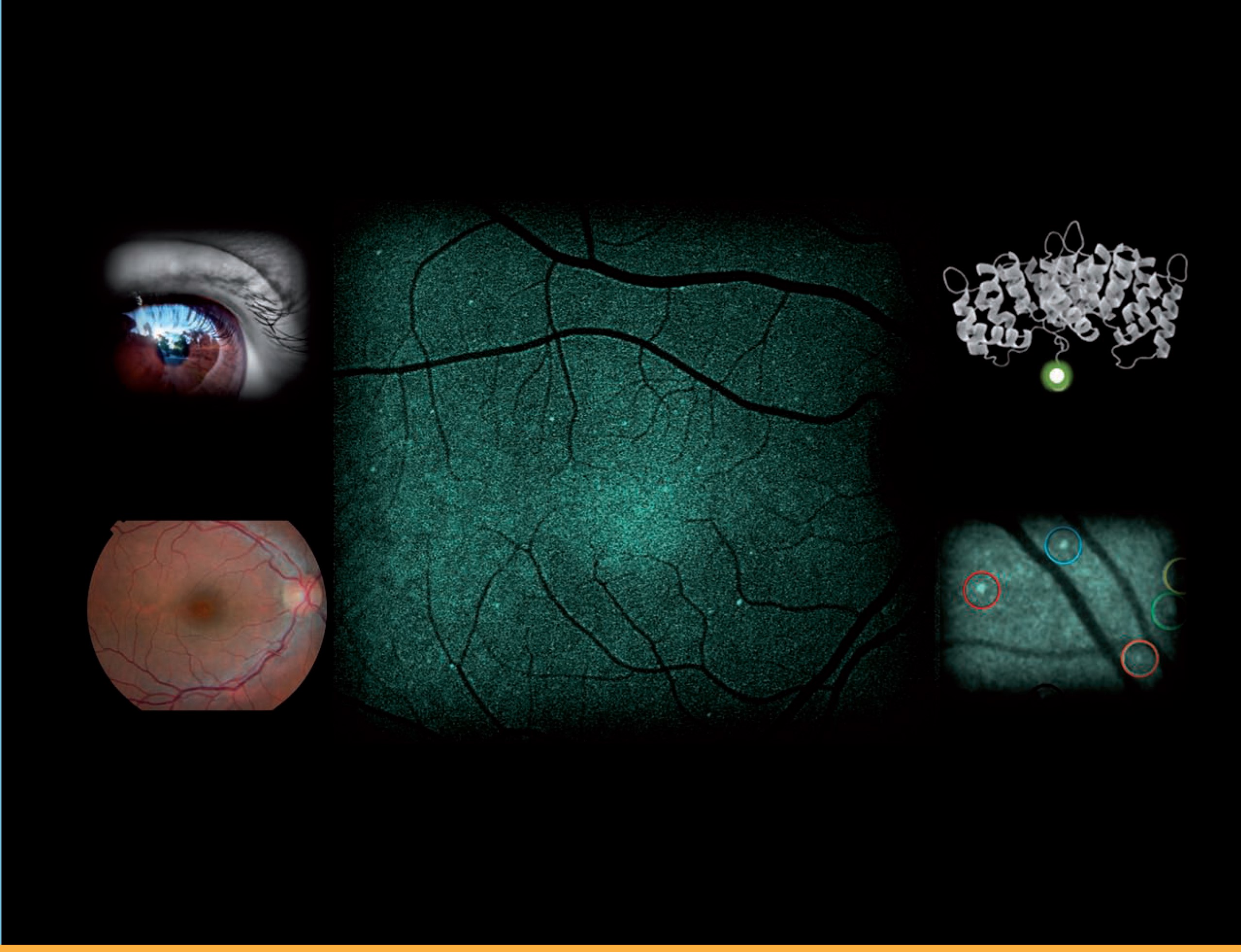
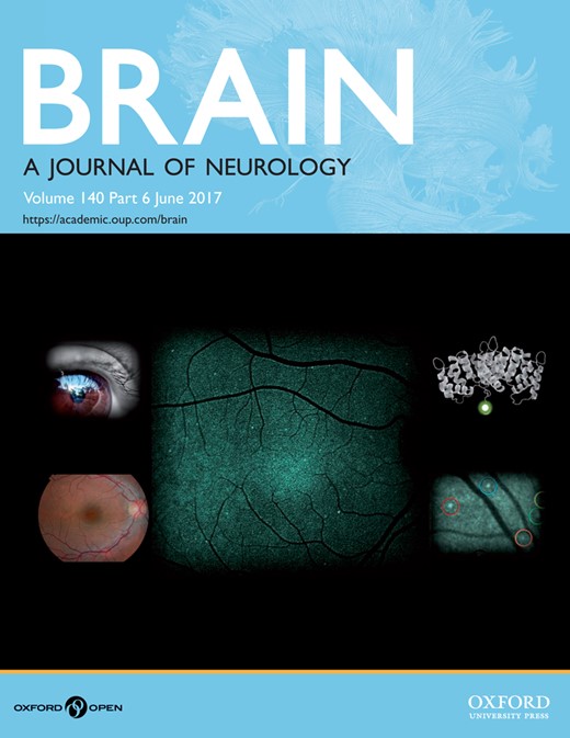
Cover image: Intravenous injection of a fluorescently-tagged annexin molecule enables identification of individual apoptosing neuronal cells in the retina. A novel in vivo technique, DARC, was used to label these ‘sick’ retinal nerve cells. From Cordeiro et al. Real-time imaging of single neuronal cell apoptosis in patients with glaucoma. Pp. 1757–1767.
Volume 140, Issue 6, June 2017
Editorial
Transdiagnostic neurology: neuropsychiatric symptoms in neurodegenerative diseases
Scientific Commentaries
Holes in the leaky migraine blood–brain barrier hypothesis?
This scientific commentary refers to ‘Increased brainstem perfusion, but no blood-brain barrier disruption, during attacks of migraine with aura’, by Hougaard et al.. (doi:).
Cortical areas needed for choosing actions based on desires
This scientific commentary refers to ‘Selective impairment of goal-directed decision-making following lesions to the human ventromedial prefrontal cortex’, by Reber et al. 2017 (doi:).
In vivo imaging of retinal neurodegeneration at the single cell level in humans
This scientific commentary refers to ‘Real-time imaging of single neuronal cell apoptosis in patients with glaucoma’, by Cordeiro et al. (doi:).
Drug repurposing to target proteostasis and prevent neurodegeneration: accelerating translational efforts
This scientific commentary refers to ‘Repurposed drugs targeting eIF2α-P-mediated translational repression prevent neurodegeneration in mice’, by Halliday et al.. (doi:).
Review Article
Glioblastoma-associated microglia and macrophages: targets for therapies to improve prognosis

Glioblastoma-associated microglia and macrophages initially participate in tumour surveillance but are then subverted to assume tumour-promoting roles. Poon et al. review the biology of these innate immune cells and the model systems used to study them, as well as recent advances in immunotherapies targeting these cells in glioblastoma.
Original Articles
Hypomorphic mutations in POLR3A are a frequent cause of sporadic and recessive spastic ataxia

The genetic basis of many spastic ataxias remains unknown. Minnerop et al. identify intronic mutations in POLR3A as a frequent cause of recessive spastic ataxia. Surprisingly, >80% of carriers have the same c.1909+22G>A mutation. The mutation causes a homogeneous phenotype without overt leukodystrophy, previously considered the unifying feature of POLR3A-disease.
A panel study on patients with dominant cerebellar ataxia highlights the frequency of channelopathies

Polyglutamine expansions are the most frequent cause of autosomal dominant cerebellar ataxias. Using a panel sequencing approach in a cohort of 412 patients, Coutelier et al. identify mutations in ion channels as the next most frequent cause. They describe genotype-phenotype correlations relevant for genetic counselling and with possible prognostic value.
ATAD3 gene cluster deletions cause cerebellar dysfunction associated with altered mitochondrial DNA and cholesterol metabolism

Mitochondrial DNA dysfunction causes a range of neurological diseases. Desai, Frazier et al. show that deletions in the ATAD3 gene cluster create chimeric proteins that are associated with cerebellar defects, mitochondrial DNA disorganisation and perturbed cholesterol homeostasis. The findings link mitochondrial DNA, cholesterol, and brain development and function.
A comprehensive analysis of rare genetic variation in amyotrophic lateral sclerosis in the UK

The genetic landscape of amyotrophic lateral sclerosis (ALS) is poorly understood. By examining known ALS-associated genes in a large cohort, Morgan, Shatunov et al. report an increase in mutations within the untranslated prime regions of the genes and a greater than expected number of patients with multiple potentially pathogenic variants.
A novel cortical target to enhance hand motor output in humans with spinal cord injury

Increased brainstem perfusion, but no blood-brain barrier disruption, during attacks of migraine with aura

See Moskowitz (doi:) for a scientific commentary on this article.
Animal studies suggest that cortical spreading depression — the likely mechanism of migraine aura — disrupts the blood-brain barrier, and thereby leads to migraine headache. However, using an advanced MRI technique in patients, Hougaard et al. demonstrate that headache following spontaneous aura is associated with brainstem hyperperfusion while the blood-brain barrier remains intact.
Inhibition of the P2X7–PANX1 complex suppresses spreading depolarization and neuroinflammation

Spreading depression (SD) is a wave of neuroglial depolarization. Chen et al. show that inhibition of the neuronal P2X7-Panx1 pore-complex suppresses SD, as well as SD-induced neuroinflammation and activation of trigeminal neurons. Inhibition of P2X7-Panx1 may therefore be a promising target for diseases associated with SD, such as migraine.
Rare missense mutations in P2RY11 in narcolepsy with cataplexy

Narcolepsy with cataplexy (type I narcolepsy) is thought to be an autoimmune disorder but its precise pathogenesis is unknown. Degn et al. report eight rare missense mutations in P2RY11, found exclusively in affected individuals. Six of the mutations disrupt P2Y11 receptor function, implicating decreased P2Y11 signalling in the underlying aetiology.
The spectrum of REM sleep-related episodes in children with type 1 narcolepsy

Paediatric type 1 narcolepsy is characterised by cataplexy and complex movement disorders during wakefulness. Using video-polysomnography to analyse motor events during sleep, Antelmi et al. show that up to one third of patients also display complex motor behaviours during REM sleep. Night-time motor dyscontrol correlates with severe day-time cataplectic features.
Crowdsourcing seizure detection: algorithm development and validation on human implanted device recordings

Automated seizure detection algorithms are needed for both basic research and epilepsy therapy. Baldassano et al. present results from an open competition to crowdsource algorithm development, and demonstrate the efficacy of these algorithms on out-of-sample human recordings. The algorithms offer translational utility and set a reproducible benchmark for seizure detection.
Imaging blood–brain barrier dysfunction as a biomarker for epileptogenesis

A biomarker for the identification of patients at high-risk for post-injury epilepsy is urgently needed. Bar-Klein et al. report that in the rat status-epilepticus model, region-specific blood-brain barrier pathology detected by contrast-enhanced MRI serves as a diagnostic, predictive and pharmacodynamic epilepsy biomarker.
Lesion mapping of stroke-related erectile dysfunction

Acute ischaemic stroke in brain areas contributing to male sexual arousal may impair erectile function. Using a voxel-wise analysis, Winder et al. report that deterioration of erectile function after stroke correlates with ischaemic lesions in the right occipito-parietal and thalamic areas, and left insular and temporo-parietal areas.
Right hemisphere structural adaptation and changing language skills years after left hemisphere stroke

Language difficulties after stroke are commonly thought to stabilise within a year. Hope et al. report surprising evidence to the contrary, showing that the language skills of patients with post-stroke aphasia continue to change even years after stroke. The changes are associated with structural adaptation in the intact right hemisphere.
Using transcranial magnetic stimulation of the undamaged brain to identify lesion sites that predict language outcome after stroke

Predicting how brain damage will affect cognition is notoriously challenging. By using findings from non-invasive brain stimulation studies of healthy subjects to guide lesion-deficit mapping, Lorca-Puls et al. identify two lesion sites, in frontal and parietal areas, that consistently cause persistent language difficulties after stroke.
Selective impairment of goal-directed decision-making following lesions to the human ventromedial prefrontal cortex

See Manohar and Akam (doi:) for a scientific commentary on this article.
Using a food reward devaluation procedure, Reber et al. show that patients with ventromedial prefrontal cortex lesions are selectively impaired in instrumental choices following satiation. This argues for a role for the human vmPFC in goal-directed decision-making, while showing that it is not necessary for hedonic experience of outcome value.
Real-time imaging of single neuronal cell apoptosis in patients with glaucoma

See Herms and Schön (doi:) for a scientific commentary on this article.
Glaucoma is often diagnosed late when vision loss has already occurred. Cordeiro et al. report a new fluorescent marker for retinal imaging that can safely visualise real-time in vivo neuronal apoptosis in patients. Increased labelling is observed in patients with progressive neurodegenerative disease compared to healthy controls.
Repurposed drugs targeting eIF2α-P-mediated translational repression prevent neurodegeneration in mice

See Mercado and Hetz (doi:) for a scientific commentary on this article.
Signalling through the PERK/eIF2α-P branch of the Unfolded Protein Response is increased in many neurodegenerative diseases. Halliday et al. identify two safe compounds – one licensed – that act on this pathway and are neuroprotective in mice with neurodegeneration. These drugs can now be repurposed in clinical trials for the treatment of dementia.
Cognitive reserve and TMEM106B genotype modulate brain damage in presymptomatic frontotemporal dementia: a GENFI study

Frontotemporal dementia (FTD) shows substantial phenotypic variability. In a multicentre study, Premi et al. explore the effect of cognitive reserve and TMEM106B genotype in modulating grey matter volume in presymptomatic FTD. Environmental as well as genetic factors affect rates of brain atrophy, suggesting a possible strategy for delaying disease onset.
Apathy and impulsivity in frontotemporal lobar degeneration syndromes

Lansdall et al. reveal the structural brain changes leading to different types of apathy and impulsivity, across the spectrum of disorders caused by frontotemporal lobar degeneration, including frontotemporal dementia, progressive supranuclear palsy and corticobasal syndrome. The positive correlation between apathy and impulsivity indicates the need for a unified therapeutic strategy.
Dorsal column
From the Archive
Progressive atrophy of the globus pallidus (primary atrophy of the pallidal system). A system disease of the paralysis agitans type, characterized by atrophy of the motor cells of the corpus striatum. A contribution to the function of the corpus striatum. By J. Ramsay Hunt MD, New York. Brain 1917; 40: 58–148

Book Review
Dramatic science or scientific drama?


