 Musculoskeletal Imaging Volume 2: Metabolic, Infectious, and Congenital Diseases; Internal Derangement of the Joints; and Arthrography and Ultrasound
Contents
-
-
-
-
-
-
-
-
-
Introduction Introduction
-
Pathophysiology Pathophysiology
-
Clinical Findings Clinical Findings
-
Imaging Strategy Imaging Strategy
-
Imaging Findings Imaging Findings
-
Radiography Radiography
-
Computed Tomography Computed Tomography
-
Magnetic Resonance Imaging Magnetic Resonance Imaging
-
Nuclear Imaging Nuclear Imaging
-
-
Differential Diagnosis Differential Diagnosis
-
Treatment Options Treatment Options
-
Recommended Reading Recommended Reading
-
References References
-
-
-
-
-
-
-
-
-
-
-
-
-
-
-
-
-
-
-
86 Osteomyelitis of the Long and Flat Bones
-
Published:April 2019
Cite
Abstract
Chapter 86 highlights the imaging manifestations of osteomyelitis (OM) involving the long and flat bones. OM refers to inflammation of the bone and bone marrow caused by underlying infection, classically bacterial. Long and flat bone OM can occur either because of hematogenous spread, direct inoculation or from a contiguous source of infection. The severity depends on the factors such as organism isolated, pathogenesis, extent of bone involvement, duration of infection, and host factors such as age and immune status. Imaging plays a crucial role in the early diagnosis of OM with MRI being the modality of choice. Both acute and chronic forms of OM are still a big challenge to treat, even in the era of advanced antibiotics and new surgical techniques. Imaging helps in early diagnosis, which in turn helps to initiate early treatment.
Introduction
Osteomyelitis (OM) is defined as inflammation of the bone and bone marrow as a result of bacterial infection. Other less common causative agents include fungi, parasites, and viruses.
Pathophysiology
Long bone OM can occur secondary to hematogenous spread, direct inoculation into bone, or from a contiguous source of infection. Hematogenous spread is more common in children and elderly people. In adults, hematogenous OM commonly affects the spine, pelvis, and small bones. Direct inoculation usually occurs secondary to surgery or trauma. OM caused by adjacent soft tissue infection is commonly seen in patients with diabetes mellitus or in bedridden patients.
The hematogenous form is usually caused by a single pathogen, the commonest organism being Staphylococcus aureus (S. aureus) in children and adults. A new gram-negative coccobacillus organism, Kingella kingae, is now isolated with greater frequency in children. Children with sickle cell disease may be infected with Salmonella. The commonest organisms isolated in infants are S. aureus, Streptococcus agalactiae, and Escherichia coli. Increasingly, methicillin-resistant S. aureus (MRSA) is being isolated from patients with OM. In OM secondary to direct inoculation or contiguous spread, multiple organisms may be isolated.
Hematogenous OM has a predilection for the metaphyses of long bones and is attributed to the peculiar vascular pattern in this region, where terminal branches of the nutrient artery form a mesh of hairpin bends before draining into a large network of sinusoidal veins. This arrangement causes blood to flow slowly in the metaphysis, favoring bacterial colonization. In infants and adults, the metaphyseal and epiphyseal vessels communicate with each other, hence the metaphyseal infection can pass to the epiphysis. In children, however, very few vessels cross the epiphyseal plate, which act as a relative barrier to the spread of infection. A recent study has shown a higher incidence of transphyseal spread of infection than previously thought.
The acute stage of OM is characterized by BME caused by inflammatory exudates. As the infective process progresses across the medullary cavity, the increased intraosseous pressure causes pus to track within Volkmann canals and spread subsequently into the subperiosteal space, causing periosteal stripping. If untreated, the infected bone undergoes devascularization secondary to vascular stasis and small-vessel thrombosis, resulting in a necrotic bone fragment known as sequestrum. If the infective process is aborted at this stage by appropriate treatment, resolution and healing occurs. If the infection persists beyond 6 weeks, chronic OM is established, characterized by an involucrum, which is an area of periosteal new bone encasing the sequestrum. The involucrum may develop perforations, termed cloaca, through which the pus may track into the surrounding soft tissue and eventually to the skin surface via a sinus tract.
Clinical Findings
A high index of clinical suspicion, together with laboratory and imaging findings, is required to diagnose OM. The clinical symptoms are usually nonspecific and include fever, chills, swelling, erythema, and pain over the involved bone. Children are usually irritable and lethargic. In cases of direct inoculation or contiguous spread of disease, patients may present with focal bone pain, erythema, swelling, and pus drainage around the area of surgery, trauma, or wound infection. Laboratory investigations should include complete blood count, ESR, CRP, cultures, and Gram stain. Cultures of blood and material obtained by needle aspiration of the involved bone are necessary to identify the causative organism and yield positive findings in up to 87% of cases. Bone biopsies usually taken at the time of debridement should undergo histopathologic examination and culture and sensitivity.
Imaging Strategy
The imaging modalities frequently employed to diagnose OM are conventional radiography, radionuclide scanning, CT, and MRI. In all patients with suspected OM, evaluation usually begins with radiographs. Figures 86.1–86.5 demonstrate radiographic, CT, MRI, and radionuclide imaging findings of acute and chronic OM.

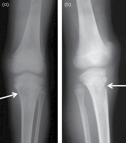
Acute OM of the left tibia in an 18-year-old man. (A) AP and (B) oblique radiographs of the tibia and fibula show permeative bone destruction (arrows) involving the proximal metaphysis of the left tibia. The lesion has wide zone of transition. There is cortical break noted anteriorly with adjacent soft tissue edema.
MRI is the modality of choice in imaging OM because of its high spatial resolution, multiplanar ability, and better soft tissue contrast. In OM, the specificity and sensitivity of MRI ranges from 50% to 90% and 60% to 100%, respectively. MRI can detect changes as early as 3 to 5 days after the infective process begins. MRI protocol should include T1W and T2W sequences supplemented by T2W FS or STIR sequences. IV Gd-based T1W FS postcontrast sequences are helpful in depicting nonenhancing intraosseous, subperiosteal, and soft tissue fluid collections and areas of tissue necrosis, rim-enhancing abscesses, and enhancing phlegmons.
In the acute stage, when the radiographs are not of much help, radionuclide imaging may be useful for the early detection of OM. It can detect OM as early as 48 hours after the onset of infection. Additionally, bone scan is helpful in detecting multifocal OM.
Imaging Findings
Radiography
In the early stages of OM, radiographs may be normal and changes are usually visible approximately 2 to 3 weeks after the onset of infection.
Early radiographic findings include soft tissue swelling, subcutaneous edema, and blurring of the soft tissue planes.
In OM caused by contiguous spread of infection, the earliest radiographic finding is usually soft tissue edema and periosteal reaction that closely resembles posttraumatic periosteal reaction and periosteal reaction caused by chronic venous stasis.
As disease progresses, osseous findings such as osteoporosis, permeative bone erosion, and trabecular destruction caused by infection in the medullary space are seen (Figure 86.1).
As infection spreads outward across the haversian and Volkmann canals, there is increased osteolysis and cortical erosion (Figure 86.1). This is followed by periostitis, caused by lifting of the periosteum by the subperiosteal abscess.
In adults, the periosteum is attached firmly to the cortex, ensuring good cortical blood supply and resisting periosteal elevation and new bone formation.
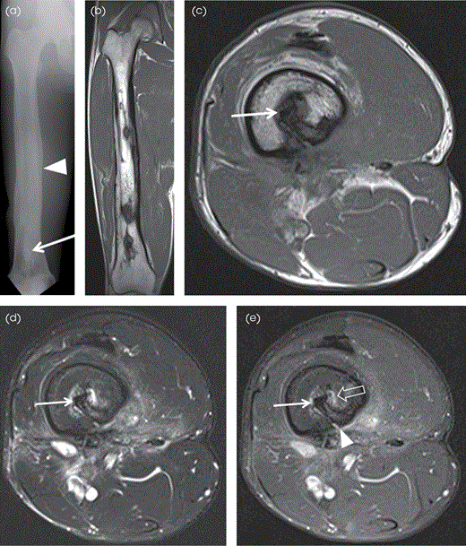
In late stage or chronic stage of OM, radiographs usually reveal bone expansion and remodeling, cortical thickening, and may show sequestrum, involucrum, and cloaca (Figure 86.2A).
A sequestrum is seen as a sclerotic bone fragment within a lucent lesion.
An involucrum is thick periosteal new bone surrounding the sequestrum.
Cloaca appears as a lucent defect within the cortical bone.
Sequestrum and involucrum are unusual findings in adults compared to the pediatric population.
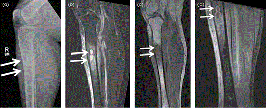
Some patients, particularly children, may develop an intraosseous abscess known as a Brodie abscess in the subacute or chronic stages of OM (duration of at least 6 weeks). These are seen as well-defined round to oval lucent lesions with dense surrounding sclerosis and associated periosteal reaction (Figure 86.3A). Brodie abscess commonly involves the metaphysis of the long bones, particularly the distal or proximal portions of the tibia.

Chronic OM of the right femur in a 32-year-old man. (A) AP radiograph of the right femur shows cortical thickening, osseous expansion and bony remodeling, sclerotic changes, sequestrum (arrow), and involucrum (arrowhead). Coronal (B) and (C) axial T1W, (D) axial T2W FS, and (E) axial postcontrast T1W FS MR images show diffuse cortical thickening involving the right femur with medullary sequestrum (arrow), which is hypointense on all sequences, enhancing surrounding granulation tissue (open arrow) and cloaca (arrowhead), which is seen as a cortical defect.

Brodie abscess in the right tibial diaphysis in a 48-year-old woman. Lateral radiograph (A) shows a bilobed lucent lesion with a thin sclerotic rim in the proximal tibial diaphysis consistent with Brodie abscess (arrows). (B) The lesion demonstrates high signal intensity on the sagittal STIR MR image with associated low signal rim of sclerosis (arrows) and adjacent high signal BME. On the sagittal T1W MR image (C) the lesion shows central low signal with higher/intermediate signal intensity rim (penumbra sign) and surrounding low signal sclerosis (arrows) and demonstrates rim enhancement (arrows) on the postcontrast T1W FS MR image (D) with enhancing adjacent BME.
Computed Tomography
Although CT is not the modality of choice in OM, it is superior to radiographs in demonstrating the amount of bone destruction, periosteal reaction, intraosseous gas, and soft tissue extension (Figure 86.4A,B).
CT is more useful in cases with chronic OM, for identifying sequestra, involucra, and cloacae, features that are important in deciding surgical management.
Another advantage of CT is its usefulness in image-guided biopsy.

OM of the left clavicle in a 24-year-old man. (A) AP radiograph of the left shoulder shows osseous expansion of the proximal and mid clavicle with periosteal reaction (arrow). (B) Axial CT image of the left clavicle, taken in bone window, better demonstrates osteolytic and sclerotic changes with periosteal reaction (arrows). (C) Axial T2W FS MR image shows diffuse increased signal in the left clavicle with periosteal reaction (arrows) and surrounding reactive soft tissue edema. (D) Axial postcontrast T1W FS MR image shows diffuse periosteal (arrows), marrow and surrounding soft tissue enhancement in keeping with acute OM. (E) Corresponding 99mTc-MDP radionuclide bone scan shows intense increased uptake in the left clavicle (arrow), corresponding to the site affected by acute OM.
Magnetic Resonance Imaging
BME and enhancement without associated low T1 signal comprises reactive changes/osteitis and is not diagnostic of OM.
Imaging findings of acute OM are quite nonspecific. Other conditions such as noninfectious inflammation, healing fractures, bone contusion, and metastasis can also produce increased BME on T2W images similar to OM. Thus, MRI findings must be correlated with secondary imaging abnormalities and the clinical background. The secondary findings include cortical bone destruction, cellulitis, soft tissue abscess, and sinus tract.
On MRI, the normal cortical bone appears hypointense on T1W and T2W images. In case of OM with cortical erosion, there is loss of normal cortical hypointense signal.
Soft tissue and intraosseous abscesses appear as hypointense, peripherally enhancing fluid collections on postcontrast T1W FS images (Figure 86.3D).
Sinus tracts, if present, are seen as curvilinear or linear areas of increased signal on T2W images and may exhibit contrast enhancement.
In chronic stages, sequestra and involucra demonstrate decreased signal on all the pulse sequences with no contrast enhancement (Figure 86.2B–E).
The granulation tissue surrounding the sequestrum, however, displays increased signal on fluid-sensitive sequences with contrast enhancement (Figure 86.2D,E).
Brodie abscess is best seen on MRI and appears as a round, well-defined area with decreased signal on T1W images and increased signal on T2W FS/STIR images with peripheral rim enhancement on postcontrast images. It is characteristically surrounded by a band of decreased signal on T2W images caused by bone sclerosis (Figure 86.3B-D).
It may be challenging to distinguish subacute OM from tumor on MRI images. Rim of increased and/or intermediate signal on T1W images (penumbra sign) (Figure 86.3C) favors OM over tumor.
Distinguishing acute changes of Charcot arthropathy from OM in the diabetic foot may be challenging as they can share similar imaging features on MRI. Presence of adjacent sinus tracts and draining soft tissue abscess is considered helpful in diagnosis of OM. Additionally, correlation with contemporary radiographs is useful in differentiating foot fractures from OM, which may also display similar signal on MR images. It is easier to discern frequently complex anatomy of the partially amputated and deformed diabetic foot on radiographs when compared to MR images.

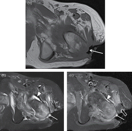
Contiguous focus OM in the left greater trochanter secondary to superficial ulcer in a 65-year-old man. (A) Axial T1W MR image shows large soft tissue defect/ulcer (arrow) by the greater trochanter with low signal in the adjacent soft tissues and in the greater trochanter. (B) Axial STIR MR image shows increased signal in the greater trochanter (arrow) and surrounding soft tissues with associated left hip joint effusion (arrowhead). (C) Corresponding axial postcontrast T1W FS MR image shows enhancement in the affected greater trochanter (arrow) and the soft tissues with enhancing synovitis (arrowhead) in the left hip. Note nonenhancing devitalized tissue (block arrow).
Nuclear Imaging
The commonly used radionuclide, technetium-99m-labeled methylene diphosphonate (99mTc-MDP) demonstrates increased tracer uptake in acute OM (Figure 86.4E). Other agents used are gallium-67 citrate, and indium-111–labeled leukocytes.
Radionuclide imaging is highly sensitive but has the disadvantage of low specificity and poor anatomical details. Consequently, it is at times difficult to differentiate between OM and other entities such as fractures, neoplasia, or arthritis.
Differential Diagnosis
Neoplasms such as osteosarcoma, Ewing sarcoma, lymphoma, and metastasis
LCH
Osteoid osteoma
Charcot arthropathy in diabetic foot
Fractures
Treatment Options
OM treatment depends on the appropriate antibiotic therapy and often requires thorough surgical debridement removing infected and necrotic tissue, adequate drainage, postdebridement dead space obliteration, and proper wound care.
If OM is secondary to host-related causes, such as diabetes mellitus, effort should be made to treat the underlying etiology and improve the host immune system.
When possible, the antibiotic treatment should be based on the culture and susceptibility results. In cases of urgent surgical debridement, before the cultures can be obtained, empirical broad-spectrum antibiotics should be administered.
Indications for surgical management include chronic OM, failed response to the antibiotic treatment, and infected surgical hardware.
Surgical management helps remove dead necrotic tissues and decreases the bacterial load.
OM is caused by hematogenous spread, direct inoculation or contiguous spread from adjacent infection.
Acute stage radiographic findings include soft tissue edema, osteoporosis, permeative bone erosion, cortical erosion, and periosteal reaction.
Sequestra, involucra, and cloacae are markers of the chronic stage.
Brodie abscess usually affects children and involves the metaphysis of the long bones.
MRI is the modality of choice because of higher sensitivity and better soft tissue contrast.
MRI protocol must include a T1W sequences together with fluid-sensitive sequences.
Postcontrast T1W FS MR images improve delineation of nonenhancing fluid collections and necrotic tissue and rim-enhancing osseous and soft tissue abscesses.
Recommended Reading
Tehranzadeh J, Wong E, Wang F, Sadighpour M.
References
1. Calhoun JH, Manring MM, Shirtliff M.
2. Gilbertson-Dahdal D, Wright JE, Krupinski E, McCurdy WE, Taljanovic MS.
3. Hatzenbuehler J, Pulling TJ.
4. Ikpeme IA, Ngim NE, Ikpeme AA.
5. McGuinness B, Wilson N, Doyle AJ.
6. Oudjhane K, Azouz EM.
7. Pineda C, Espinosa R, Pena A.
8. Renton P. Periosteal reaction, bone and joint infections. In: Sutton D, ed.
9. Sanders J, Mauffrey C.
10. Tehranzadeh J, Wong E, Wang F, Sadighpour M.
| Month: | Total Views: |
|---|---|
| October 2022 | 6 |
| December 2022 | 3 |
| January 2023 | 2 |
| February 2023 | 4 |
| March 2023 | 4 |
| April 2023 | 1 |
| May 2023 | 1 |
| June 2023 | 6 |
| July 2023 | 2 |
| August 2023 | 2 |
| September 2023 | 2 |
| October 2023 | 2 |
| November 2023 | 2 |
| December 2023 | 2 |
| January 2024 | 1 |
| February 2024 | 1 |
| March 2024 | 2 |
| April 2024 | 3 |
| May 2024 | 7 |
| June 2024 | 3 |
| July 2024 | 3 |
| August 2024 | 1 |
