


Abstract
Sepsis-induced acute kidney injury (SI-AKI) is a severe complication of critical patients with high morbidity and mortality. Apoptosis, pyroptosis, and necroptosis, regulated by a common pathway - PANoptosis, play essential roles in SI-AKI without the clarified mechanisms.
We first established the SI-AKI model by intraperitoneal injection of the lipopolysaccharide (LPS) (O111:B4) in the C57B/L6J mice with increased Serum creatinine (Scr), blood urea nitrogen (BUN), and kidney injury biomarkers (NGAL, KIM-1) in renal tissue. The expression of crucial molecules in PANoptosis were examined by quantitative real-time PCR (qPCR) and Western blotting. RIPK1 inhibitor-necrostatin-1(Nec-1) (1.65mg/kg) and pan-caspase inhibitor- Emricasan (10mg/kg) were further employed to mice with LPS. Potential interacting proteins of panoptosome were detected by co-immunoprecipitation. We also analyzed the methylation level of ZBP1 in cell-free DNA among sepsis-non-AKI and SI-AKI patients.
The expression of crucial molecules in PANoptosis, including IL-1β(0.82 vs. 0.35, P = 0.003), mixed lineage kinase domain-like (MLKL) (0.67 vs. 0.41, P = 0.014), protein kinase 1 (RIPK1) (1.07 vs. 0.72, P = 0.002), cleaved-caspase 3 (1.13 vs. 0.59, P < 0.001), and FAS-associated death domain protein (FADD) (0.78 vs. 0.43 P < 0.001) elevated in the SI-AKI group. Z-DNA binding protein 1 (ZBP1), a typical sensor of PANoptosis, is dramatically activated in the SI-AKI mice. Co-immunoprecipitation showed that caspase 8, ZBP1, peptidoglycan recognition protein 1 (pglyrp1), and RIPK1 are potential components of PANoptosome. Nec-1 and Emricasan could attenuate the LPS-induced increases in the concentrations of BUN and inflammatory cytokine (TNF-a and IL-6), as well as the methylation level of ZBP1 in cell-free DNA decreased in SI-AKI patients verse sepsis-none-AKI (P = 1.60E-06).
ZBP1 exacerbated SI-AKI via activating PANoptosis with the potencial PANoptosome of ZBP1, caspase 8, pglyrp1, and RIPK1.
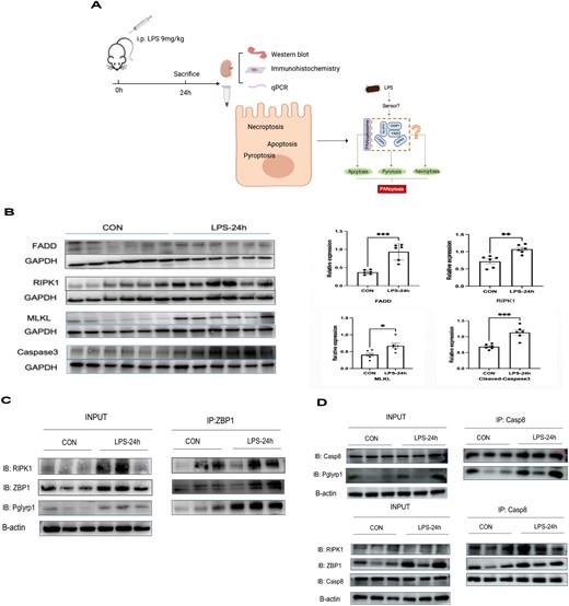
A: The overview of the study design. B. Crucial molecules in PANoptosis elevated in SI-AKI. C. The potential factors of the PANoptosome. Data are means ± SEM of 3 independent experiments (n = 6 mice/group). *P < 0.05, **P < 0.01, ***P < 0.001.


Comments