


Abstract
Pericytes are mesenchyme-derived perivascular cells attached to the abluminal surface of capillaries as well as the major origin of myofibroblasts in the animal model of unilateral ureteral obstruction (UUO) and ischemia-reperfusion injury (IRI). Although previous studies reported that myofibroblasts may be beneficial during acute kidney injury (AKI) via growth factor secretion, reconstitution and stabilization of the collagen framework for tubular cell regeneration, lack of solid evidence and definite mechanism make the role of pericyte/myofibroblast in AKI remain controversial.
To clarify this, we used platelet-derived growth factor receptor β (PDGFRβ)-specific antibody following AKI in C57BL/6 mice to inhibit pericytes. In the meantime, We used Gli1-CreERT2;Cxcl12fl/fl genetically modified mice to conditionally knockout CXCL12 specifically in pericytes before IRI.
The expression of PDGFRβ on pericytes increased after IRI. In group of administration of PDGFRβ antibody, proliferation and transition to myofibroblasts of pericytes were inhibited. Inflammation became worse and renal recovery was impeded after AKI. The chemokine stromal cell-derived factor-1 (SDF-1) which is also known as the C-X-C-type chemokine CXCL12 decreased as well. Furthermore, we demonstrated that CXCL12 was secreted mainly by pericytes and its receptor CXCR4 was expressed on tubular cell during IRI by in situ hybridization. When knockout of CXCL12 was performed before IRI, the renal microvascular rarefaction was noted after AKI and the recovery of renal function was impaired. Renal tubular cell proliferation decreased and apoptosis increased as well.
In summary, these findings demonstrated that pericytes contribute to renal repair after IRI through CXCL12 secretion.
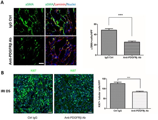
Anti-PDGFRβ Ab inhibited pericyte–myofibroblast transition and tubular regeneration after IRI. (A) Representative images showed aSMA+ cells of the kidneys at day 5 after IRI with or without anti-PDGFRβ antibody administration. Bar chart showed the numbers of aSMA+ cells. (B) Representative images showed Ki67+ cells of the kidneys at day 5 after IRI with or without anti-PDGFRβ antibody administration. Bar chart showed the numbers of Ki67+ cells. Scale bar, 50 μm. Original magnification × 200. n = 5 for each group. Data were expressed as the mean ± SEM. *P < 0.05, **P < 0.01, ***P < 0.001 by Student's t-test.


Comments