


 https://orcid.org/0000-0002-0031-2392

Abstract
Candida auris is an emerging multidrug-resistant fungal pathogen that uniquely colonizes the skin. The factors regulating C auris colonization are poorly understood. In this study, we examined the role of galectin-3 during C auris skin infection. We found that deletion of galectin-3 enhances the antifungal activity of neutrophils and increases resistance to C auris skin infection. Mechanistically, galectin-3 negatively regulates GLUT4 expression, glucose uptake, reactive oxygen species production, and fungal killing by neutrophils. Collectively, our findings uncover a detrimental role for galectin-3 in neutrophil-mediated defense against C auris.
Candida auris, a multidrug-resistant fungal pathogen, colonizes the human skin distinctively and long-term, leading to nosocomial transmission and outbreaks of systemic fungal infections. The US Centers for Disease Control and Prevention and the World Health Organization have designated C auris an urgent concern and placed it under a critical priority fungal infections group [1]. Most C auris isolates exhibit resistance to several Food and Drug Administration–approved antifungal drugs; therefore, understanding the mechanistic basis of skin colonization by C auris is critical to developing novel host-directed antifungal therapeutics to prevent and treat this emerging fungal pathogen. Although antifungal host defense mechanisms against C albicans are well studied [2], much less is known about the host immune responses that regulate C auris skin colonization.
Galectin-3 is a β-galactoside–binding lectin implicated in infection, inflammation, and disease states [3]. Galectin-3 is mainly found in the cytoplasm and can be translocated to the nucleus or secreted onto the cell surface. Additionally, it is released into bodily fluids, including serum and urine [3]. Galectin-3 is expressed and secreted by almost all cell types, including dendritic cells, eosinophils, mast cells, natural killer cells, endothelial cells, and epithelial cells [3]. The role of galectin-3 during invasive candidiasis has been less clear, as protective and detrimental roles have been reported [4–6]. In this study, we examined how galectin-3 affects host defense during C auris skin infection. Our study uncovers a previously unknown detrimental role of galectin-3 in modulating the antifungal activity of neutrophils by regulating glucose transporters (GLUTs), glucose uptake, reactive oxygen species (ROS) production, and fungal killing.
METHODS
Mice
C57BL/6 and galectin-3 knockout C57BL/6J strains were obtained from Jackson Laboratory and bred in centrally managed animal facilities at Purdue University.
Ethics Statement
This study followed the Guide for the Care and Use of Laboratory Animals (National Institutes of Health). Animal studies and experimental protocols were approved by the Purdue University Institutional Animal Care and Use Committee (protocol 2110002211).
Fungal Culture
The C auris AR0387 strain used in the study was procured from the Centers for Disease Control and Prevention’s AR Isolate Bank. For neutrophil ex vivo assays, C auris was preincubated with 10% mouse serum (Invitrogen) at 37 °C for 30 minutes.
C auris Murine Skin Infection
Intradermal infection was performed as previously described [7].
Isolation and Staining of Immune Cells
Skin tissue samples were collected and processed as described. Flow cytometric data were acquired through an Attune NxT Flow Cytometer (Invitrogen) and analyzed with FlowJo software.
Neutrophil Fungal Killing
Mouse femurs and tibias were harvested, and neutrophils were isolated per the Histopaque gradient density method. Neutrophils were infected with C auris at a multiplicity of infection of 4:1. The number of live C auris was assessed by plating serial dilutions on YPD agar at 3 hours postincubation. Percentage killing was expressed as follows: [1 – (CFU [colony-forming units] of C auris in the presence of neutrophils / CFU of C auris in the absence of neutrophils)] × 100. Fold change C auris killing was expressed as percentage killing in the experimental group over percentage killing in the control group. GB1107 was added 30 minutes prior to infection and continued until termination, wherever necessary.
Macrophage Fungal Killing
Bone marrow–derived macrophages were isolated from mouse femur and tibia and seeded in 96-well plates, each containing 1 × 10⁵ cells for at least 24 hours. The number of live C auris was assessed by plating serial dilutions on YPD agar at 3 hours postincubation. Percentage killing was expressed as follows: [1 – (CFU of C auris in the presence of neutrophils / CFU of C auris cells in the absence of neutrophils)] × 100. Fold change C auris killing was expressed as percentage killing in the experimental group over percentage killing in the control group.
RNA Extraction and Quantitative Real-time Polymerase Chain Reaction
Neutrophils were seeded and infected with C auris. After 3 hours of incubation at 37 °C, cells were lysed with TRIzol. RNA was extracted from neutrophils via TRIzol, according to the manufacturer's recommendations. Quantitative real-time polymerase chain reaction was performed with SYBR Green FastMix. The expression of each gene was normalized to that of Gapdh.
ROS Measurement
Bone marrow neutrophils were incubated with C auris for 3 hours. The ROS Detection Cell-Based Assay Kit (601520; Cayman Chemicals) and Mitochondrial ROS Detection Assay Kit (701600; Cayman Chemicals) were used per the manufacturer's protocol. ROS production was measured by SpectraMax i3x (Molecular Devices). 2-Deoxy-D-glucose was added 30 minutes prior to infection and continued until termination, wherever necessary.
Glucose Uptake Measurement
Bone marrow neutrophils were seeded. The media was changed to RPMI (25mM glucose with 0.25% bovine serum albumin) for starvation. Cells were stimulated with C auris for 3 hours. During the last 2 hours of starvation, cells were incubated with recombinant galectin-3. Next, the cells were stimulated with 100nM insulin for 20 minutes in phosphate-buffered saline (PBS). The cells were washed with PBS, and 100 mL of 2-(N-[7-nitrobenz-2-oxa-1,3-diazol-4-yl] amino)-2-deoxy-D-glucose (100 mM in PBS) was added. Cells were incubated at 37 °C for another 30 minutes. The glucose uptake by neutrophils was detected by SpectraMax i3x.
NADPH Assay
Intracellular NADPH was measured by the NADP+/NADPH Assay Kit (HY-K1501; MCE).
Statistical Analysis
All data are presented as mean (SEM). The data were statistically analyzed by the Mann-Whitney U test in GraphPad Software. All experiments were repeated, and data were pooled from 3 independent experiments.
RESULTS
Deletion of Galectin-3 Renders Mice More Resistant to Intradermal C auris Infection
We performed unbiased single-cell RNA sequencing profiling of skin tissue infected with C auris to determine the cell type–specific immune responses against C auris infection [8]. Our analysis found that Lgals3 encoding for galectin-3 was significantly upregulated in neutrophils but not macrophages within the C auris–infected skin tissue as compared with uninfected mice (Figure 1A). To determine whether galectin-3 is involved in host defense against C auris skin infection, we infected wild type (WT) and gal3−/− mice with C auris and compared their skin fungal burdens (Figure 1B). Surprisingly, we found that gal3−/− mice had significantly decreased skin fungal burden vs WT mice infected with C auris. On day 3 postinfection, the mean (SEM) fungal load in skin tissue of C auris–infected WT mice was 7.6 (0.68) log10 CFU/g of tissue, whereas that of gal3−/− mice was 7.19 (0.30) log10 CFU/g of tissue, corresponding to an approximately 60% reduction of C auris burden in gal3−/− mice as compared with WT mice.
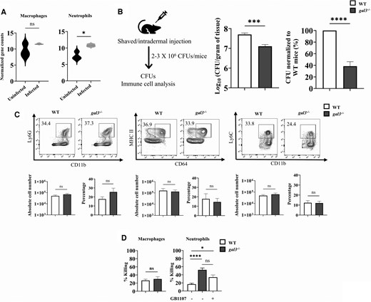
Galectin-3 is detrimental during cutaneous Candida auris infection. A, Violin plots indicate the normalized gene counts of Lgals3 in uninfected and C auris–infected skin tissue. Statistical significance was calculated by Student t test. *P < .05. B, Experimental schematic and fungal burden in the skin tissue of the indicated mice 3 days after C auris AR0387 infection. C, Representative flow cytometry plots depict the percentage and cell numbers of CD11b+ Ly6G+ neutrophils, CD64+ MHCII+ macrophages, and CD11b+ Ly6C+ monocytes in WT and gal3−/− mouse skin tissue infected with C auris AR0387. D, Macrophage and neutrophil killing activity against C auris AR0387. Data are expressed as mean (SEM) and represent 3 experiments (n = 9–12 mice/group). B–D, Statistical significance was calculated by Mann-Whitney U test. *P < .05. ***P < .001. ****P < .0001. Abbreviations: CFU, colony-forming units; gal3−/−, galectin-3 knockout; ns, not significant; WT, wild type.
Next, we examined the accumulation of myeloid phagocytes such as CD11b+ Ly6G+ neutrophils, CD11b+ MHCII+ CD64+ macrophages, CD11b+ Ly6Chi inflammatory monocytes, and CD11b+ Ly6Clow patrolling monocytes, which are essential for the host's defense against C auris skin infection [7]. We observed no significant changes in the absolute numbers of neutrophil, macrophage, and monocyte subsets between WT and gal3−/− mouse groups (Figure 1C). Since phagocyte accumulation at the site of infection was not significantly altered in the setting of galectin-3 deficiency, we next determined whether differences in the antifungal effector function of these phagocytes may contribute to the decreased fungal burden in gal3−/− mice. We isolated bone marrow–derived macrophages and neutrophils from WT and gal3−/− mice and examined their killing capacity against C auris. Although the killing activity of macrophages from WT and gal3−/− mice were similar, we found that gal3−/− neutrophils had significantly increased killing vs WT neutrophils; furthermore, addition of galectin-3 inhibitor to WT neutrophils augmented the killing capacity (Figure 1D). Collectively, these findings indicate that galectin-3 deficiency enhances the antifungal activity of neutrophils without affecting their accumulation at the site of C auris skin infection.
Galectin-3 Deficiency Augments the Fungicidal Activity of Neutrophils Through Glucose Metabolism
Next, we aimed to understand how galectin-3 regulates the antifungal activity of neutrophils against C auris. For their antimicrobial function, neutrophils require a significant amount of energy. Glucose is transported from the exterior of the cell into the cytoplasm through GLUTs and catabolized into energy, which neutrophils use to defend against fungi [9, 10]. Previous work has shown that galectin-3 binds to insulin receptors that can block the translocation of GLUT4 from the cytoplasm to the plasma membrane, thereby reducing glucose uptake in the muscle cells [11]. Also, it was recently discovered that C auris induces host metabolic stress through glucose deprivation, resulting in macrophage death [12]. On the basis of these findings, we hypothesized that galectin-3 negatively modulates GLUT4 and glucose uptake to regulate the antifungal activity of neutrophils against C auris. Therefore, we examined the expression of GLUT4 in the bone marrow–derived neutrophils isolated from WT and gal3−/− mice infected ex vivo with C auris. We found that the mRNA level of Slc2a4 transcript, which encodes for GLUT4, was significantly upregulated in gal3−/− relative to WT neutrophils from mice infected with C auris (Figure 2A). GLUT4 is an insulin-sensitive receptor that engages in insulin-stimulated glucose uptake [11, 13]. Therefore, we examined the expression of insulin receptor (Insr) but did not observe significant differences in the expression of Insr between WT and gal3−/− mice. Next, we evaluated glucose uptake and ROS activity in neutrophils from WT and gal3−/− mice. In comparison with WT neutrophils infected with C auris, we observed that glucose uptake and ROS production were elevated in gal3−/− neutrophils (Figure 2B). Furthermore, the addition of recombinant galectin-3 to neutrophils from gal3−/− mice decreased glucose uptake and ROS levels. To pinpoint the source of ROS, we examined the mitochondrial ROS levels and intracellular NADPH levels in WT and gal3−/− neutrophils. We found that gal3−/− neutrophils had higher NADPH levels, but the mitochondrial ROS was not altered after C auris infection (Figure 2C). In addition, 2-deoxy-D-glucose, which decreases glycolysis, diminished the NADPH level in gal3−/− neutrophils. To corroborate our findings with gal3−/− neutrophils, we exogenously added recombinant galectin-3 to check whether the killing capacity was restored to WT levels. Indeed, the addition of galectin-3 hampered the fungal killing in gal3−/− neutrophils. Collectively, our findings indicate that galectin-3 negatively regulates GLUT4 expression, glucose uptake, ROS production, and fungal killing (Figure 2D), thereby adversely affecting neutrophil effector functions and fungal control during cutaneous C auris infection.
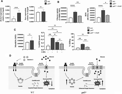
Galectin-3 negatively regulates GLUT4 expression, ROS production, and fungal killing in neutrophils. A, mRNA levels of Slc2a4 and Insr in bone marrow–derived neutrophils from WT and gal3−/− mice infected with Candida auris AR0387. B, Insulin-stimulated glucose uptake and ROS production in bone marrow–derived neutrophils of WT and gal3−/− mice in the presence or absence of exogenous recombinant galectin-3. C, Mitochondrial ROS, intracellular NADPH levels, and neutrophil killing activity in bone marrow–derived neutrophils of WT and gal3−/− mice. D, A proposed model of the role of galectin-3 in C auris clearance from murine skin. Data are expressed as mean (SEM) and represent 3 experiments (n = 3 mice/group). Statistical significance was calculated by the Mann-Whitney U test. *P < .05. **P < .01. ***P < .001. Abbreviations: gal3−/−, galectin-3 knockout; rGal3, recombinant galectin-3; ROS, reactive oxygen species; WT, wild type.
DISCUSSION
This study shows that galectin-3 deficiency enhances the antifungal activity of neutrophils and increases resistance to C auris skin infection. Galectin-3 negatively regulates GLUT4 expression, glucose uptake, and ROS production, as neutrophils from gal3−/− mice enhanced fungal killing as compared with WT mice. Although our findings suggest that galectin-3 regulates GLUT4-mediated ROS production, it is possible that it can regulate antifungal activity of neutrophils through other mechanisms. In a prior study, gal3−/− neutrophils had greater Syk activation and enhanced neutrophil function, and gal3−/− mice had reduced systemic C albicans infection [4]. In addition, dendritic cells from gal3−/− mice enhanced IL-23 secretion and decreased the susceptibility to Histoplasma capsulatum infection [14].
GLUT1 deficiency in neutrophils has been shown to exacerbate susceptibility to disseminated C albicans infection [9]. Our study mechanistically links galectin-3 to C auris infection through GLUT4 and uncovers a novel detrimental role for galectin-3 in neutrophils in the setting of cutaneous C auris infection, highlighting the potential translational implications of modulating galectin-3 to treat cutaneous fungal infections. However, as a therapeutic target against C auris infection, galectin-3 requires further research as inhibition of galectin-3 compromises the immune system and impairs immune surveillance mechanisms [15]. A combination therapy of antifungal drugs and galectin-3 inhibitors might be a novel approach to treat C auris skin infection that needs further investigation.
Note
Financial support. This study was supported by the National Institute of Allergy and Infectious Diseases, National Institutes of Health (1R01AI177604 to S. T.); and the Division of Intramural Research, National Institute of Allergy and Infectious Diseases, National Institutes of Health (ZIA AI001175 to M. S. L.).
References
Author notes
Potential conflicts of interest. All authors: No reported conflicts. All authors have submitted the ICMJE Form for Disclosure of Potential Conflicts of Interest. Conflicts that the editors consider relevant to the content of the manuscript have been disclosed.


