


 https://orcid.org/0000-0003-4444-508X

Abstract
Background
Secreted chorismate mutase (S-CM) is known to act as a virulence factor in mycobacterial infections by inhibiting macrophage apoptosis. This study aims to investigate genetic variations in S-CM among Mycobacterium intracellulare strains and evaluate their influence on the clinical progression of M intracellulare pulmonary disease (PD).
Methods
Patients diagnosed with M intracellulare PD who received treatment between 1 January 2020 and 31 December 2023 at Seoul National University Hospital were included. Clinical isolates collected at treatment initiation were assessed for subspecies classification and S-CM genetic variations. Treatment outcomes were analyzed based on subspecies and S-CM mutation status. J774A.1 murine macrophages were infected with type strains (ATCC13950T) carrying S-CM expression plasmids and clinical isolates. Macrophage apoptosis and bacterial colony-forming units (CFUs) were quantified.
Results
Among the 118 isolates, 57 were identified as M intracellulare subspecies intracellulare (typical M intracellulare [TMI]), 53 as Mycobacterium paraintracellulare, and 8 as other subspecies. A C276A mutation causing S-CM truncation was detected in 24 isolates (20.3%), all belonging to TMI. TMI strains with S-CM truncation demonstrated a higher rate of culture conversion than those with intact S-CM (adjusted hazard ratio, 2.17 [95% confidence interval, 1.08–4.34]; P = .029). In murine macrophages, TMI strains with truncated S-CM induced higher levels of apoptosis and lower CFUs, whereas expressing intact S-CM in a type strain with naturally truncated S-CM reduced apoptosis and increased bacterial burden.
Conclusions
S-CM truncation in M intracellulare enhances macrophage apoptosis, yielding reduced bacterial burden, decreased pathogenicity, and improved treatment responses.
Nontuberculous mycobacteria (NTM) comprises >190 species of mycobacteria distinct from Mycobacterium tuberculosis and Mycobacterium leprae [1]. NTM can cause chronic human infections, frequently manifesting as pulmonary disease (PD) [1]. In South Korea, the prevalence of NTM-PD has increased nearly 5-fold over the past decade, increasing from 11.4 cases per 100 000 population in 2010 to 56.7 cases in 2021 [2]. Among NTM species, M intracellulare is the leading cause of NTM-PD in South Korea, accounting for approximately 50% of cases [3].
Despite its growing epidemiological significance, the pathogenicity of M intracellulare remains controversial. Data from South Korea indicate that M intracellulare causes severe disease and is associated with higher mortality rates than Mycobacterium avium [4, 5]. Conversely, a Japanese study reported more favorable treatment outcomes for M intracellulare PD [6], whereas a study from the United States found no significant differences in virulence between M intracellulare and M avium [7]. These contrasting findings suggest that the pathogenicity of M intracellulare may vary among strains. Inflammation severity and disease progression have been shown to differ by strain and genotype of M intracellulare [8].
Recent findings suggest that secreted chorismate mutase (S-CM) functions as a virulence factor in mycobacterial infections [9, 10]. Chorismate mutase, which catalyzes the conversion of chorismate to prephenate in the shikimate pathway, exists in intracellular and secreted forms [11]. The secreted form inhibits macrophage apoptosis during Mycobacterium bovis infection, thereby enhancing bacterial survival [9]. However, the role of S-CM in NTM infections, particularly in those caused by M intracellulare, remains unclear.
We hypothesized that M intracellulare strains exhibit genetic variations in the orthologue of the Rv1885c gene (hereafter referred to as scmA), which encodes S-CM, and that these variations contribute to differences in pathogenicity. By performing genotypic analysis, we aimed to assess whether this diversity influences the clinical progression of M intracellulare PD. Additionally, we tried to validate these findings using a macrophage infection model to confirm the impact of genetic variation on pathogenicity. Therefore, this study aims to determine the potential of S-CM as a novel prognostic factor in M intracellulare PD.
MATERIALS AND METHODS
The study complied with the amended Declaration of Helsinki. The protocol was approved by the Institutional Review Board of Seoul National University Hospital (IRB number 2402-090-1511), and informed consent was waived.
Study Population and Data Collection
Patients who underwent treatment at Seoul National University Hospital between 1 January 2020 and 31 December 2023 were included. All patients met the diagnostic criteria for M intracellulare PD [1]. Treatment initiation was determined by clinical judgment of attending physicians, and all patients received guideline-based therapy [1]. Data including radiographic findings, mycobacterial cultures, drug susceptibility results, and the drugs initially administered within 1 month of treatment initiation were collected. Culture conversion and microbiological cure were determined according to the NTM-NET consensus statement [12]. Culture conversion was defined as achieving at least 3 consecutive negative mycobacterial cultures from respiratory samples, collected at intervals of at least 4 weeks. Microbiological cure was defined as maintaining multiple consecutive negative cultures following culture conversion, sustained throughout the treatment.
Subspecies Identification and scmA Genotyping From Clinical Isolates
For subspecies classification, concatenated sequences of the hsp65 and rpoB genes were analyzed using clinical isolates obtained within 3 months before treatment initiation. The hsp65 and rpoB sequences from 52 M intracellulare strains were retrieved through BLAST searches, and a concatenated phylogenetic tree was constructed (Supplementary Table 1) [13]. Representative strains from each node of the phylogenetic tree were selected to classify clinical isolates (Supplementary Figure 1). The concatenated hsp65 and rpoB sequences from clinical isolates were integrated into the phylogenetic tree to classify each clinical isolate. Details of the identification process are described in the Supplementary Material. Additionally, the scmA gene was examined for genetic variations (Supplementary Material). If NTM was still isolated after 3–6 months of treatment, subspecies identification and S-CM genotyping were also performed on those strains.
Bacterial Culture, Construction of Recombinant M intracellulare Strains, and Cell Infection
Clinical isolates and recombinant strains were used for cell infection. To complement the scmA gene, it was cloned into the integrative shuttle vector pMV306, a derivative of pMV361 [14, 15]. Recombinant M intracellulare ATCC 13950T strains were generated, carrying either the scmA-expressing plasmid under the control of the hsp65 promoter or an empty vector. Detailed procedures are described in the Supplementary Material. J774A.1 cells and THP-1 cells were infected at a multiplicity of infection (MOI) of 10. To determine intracellular and extracellular bacterial burdens, lysates, and supernatants from infected J774A.1 were plated onto 7H10 agar and incubated. Once colonies formed, colony-forming units (CFUs) were quantified (Supplementary Material).
Apoptosis and Cytokine Measurement
Annexin V-FITC Apoptosis Detection Kit with 7-AAD (Biolegend, 640922) and MitoSOX Red (Invitrogen, M36008) were used to analyze the apoptosis and mitochondrial reactive oxygen species (mtROS) production of J774A.1 cells. The supernatant from infected cells was used to quantify tumor necrosis factor alpha (TNF-α) levels (Supplementary Material).
Statistical Analysis
For continuous variables, comparisons between 2 groups were performed using the Student t test. For comparisons among 3 groups, a 1-way analysis of variance (ANOVA) was used for normally distributed variables, and the Kruskal-Wallis test for non–normally distributed variables. A 2-way ANOVA was employed to evaluate the effects of 2 independent variables on continuous outcomes. For categorical variables, group comparisons were conducted using the χ2 test or Fisher exact test, as appropriate. Time to culture conversion was analyzed using the Kaplan-Meier curves and compared using the log-rank test. Multivariate analysis was conducted using Cox proportional hazards regression. Hazard ratios (HRs) with 95% confidence intervals (CIs) were reported, and statistical significance was defined as a 2-sided P value of <.05. Phylogenetic analyses were performed using MEGA 11 software. Multiple sequence alignments were performed using the MUSCLE method, and phylogenetic trees were generated with branch support values determined from 1000 bootstrap replicates [16, 17]. Clinical and experimental data were analyzed using Stata version 18.0 (StataCorp, College Station, Texas, USA) and GraphPad Prism 10 (GraphPad Software, San Diego, California, USA), respectively.
RESULTS
Genotyping of Clinical Isolates
During the study period, 125 patients were treated, and their corresponding clinical isolates were included in the analysis. Seven isolates with unavailability for genotyping were excluded, and 118 isolates were analyzed. The clinical isolates were classified into M intracellulare subspecies: intracellulare (hereafter, typical M intracellulare [TMI]) (n = 57), paraintracellulare (hereafter, M paraintracellulare) (n = 53), yongonense (hereafter, M yongonense) (n = 5), and chimaera (hereafter, M chimaera) (n = 3).
Among the 118 isolates, 24 (20.3%) exhibited a C276A mutation in scmA, designated as ΔscmA1, resulting in a codon change from TAC (Tyr) to TAA (Stop codon) at the 92nd amino acid position. This mutation causes the truncation of S-CM, exclusively observed in TMI isolates. In contrast, none of the clinical isolates of M paraintracellulare, M yongonense, or M chimaera harbored nonsense mutations in scmA.
Phylogenetic analysis using concatenated sequences of 7 genes (hsp65, rpoB, scmA, cya, pta, glpK, and gnd) [18–20] in the 24 ΔscmA1 isolates demonstrated that they represented genetically distinct strains, rather than derivatives of a single strain (Supplementary Figure 2).
Clinical Presentation and Treatment Patterns by scmA Genetic Variation
Given that TMI accounted for a substantial proportion of isolates and ΔscmA1 was exclusively observed in TMI, patients were categorized into 3 groups: TMI with intact scmA (n = 33), TMI with ΔscmA1 (n = 24), and others (M paraintracellulare, M yongonense, and M chimaera with intact scmA) (n = 61).
While age, sex, and body mass index (BMI) were comparable across the groups, cavitation was most frequently observed in patients with TMI with intact scmA (n = 23/33 [69.7%]), followed by those with other subspecies (n = 31/61 [50.8%]) and TMI with ΔscmA1 (n = 9/24 [37.5%]) (P = .047) (Table 1). Macrolide resistance was identified in 3 patients: 1 with TMI with intact scmA and 2 with other subspecies. Detailed minimum inhibitory concentrations for each drug are provided in Supplementary Table 2. All patients received a macrolide-based regimen. Clofazimine administration was the highest in patients with TMI with intact scmA (n = 24/33 [72.7%]) and the lowest among those with TMI with ΔscmA1 (n = 9/24 [37.5%]) (P = .034) (Table 2).
Baseline Characteristics of Patients by Subspecies and scmA Genetic Variation
| Characteristic | TIM With Intact scmA (n = 33) | TIM With ΔscmA1 (n = 24) | Other Subspecies (n = 61) | P Value |
|---|---|---|---|---|
| Age, y, median (IQR) | 67 (59–73) | 67 (62–73) | 67 (61–74) | .974 |
| Female sex | 22 (66.7) | 16 (66.7) | 39 (63.9) | .967 |
| BMI, kg/m2, median (IQR) | 19.8 (1.8–21.5) | 20.6 (16.7–21.5) | 20.5 (19.1–22.5) | .124 |
| Former or current smoker | 7 (21.2) | 6 (25.0) | 13 (21.3) | .915 |
| Previous history of TB | 6 (18.2) | 4 (16.7) | 9 (14.8) | .945 |
| ESR, mm/h, median (IQR) | 32 (18–57) | 30 (17–68) | 31 (16–61) | .982 |
| AFB smear positivity | 12 (36.4) | 5 (20.8) | 14 (23.0) | .324 |
| Resistance to macrolide | 1 (3.0) | 0 | 2 (3.3) | .791 |
| Presence of cavity | 23 (69.7) | 9 (37.5) | 31 (50.8) | .047 |
Data are presented as No. (%) unless otherwise indicated.
Abbreviations: AFB, acid-fast bacilli; BMI, body mass index; ESR, erythrocyte sedimentation rate; IQR, interquartile range; TB, tuberculosis; TIM, typical Mycobacterium intracellulare.
Treatment Modalities and Outcomes of Patients by Subspecies and scmA Genetic Variation
| Treatment Modality and Outcome | TIM With Intact scmA (n = 33) | TIM With ΔscmA1 (n = 24) | Other Subspecies (n = 61) | P Value |
|---|---|---|---|---|
| Drugs administered | ||||
| Macrolide | 33 (100.0) | 24 (100.0) | 61 (100.0) | >.999 |
| Azithromycin | 33 (100.0) | 22 (91.7) | 60 (98.4) | .173 |
| Clarithromycin | 0 | 2 (8.3) | 1 (1.6) | .173 |
| Ethambutol | 31 (93.9) | 24 (100.0) | 60 (98.4) | .293 |
| Rifampicin | 7 (21.2) | 6 (25.0) | 23 (37.7) | .223 |
| Amikacin (intravenous) | 7 (21.23) | 4 (16.7) | 12 (19.7) | .953 |
| Amikacin (inhaled) | 4 (12.1) | 1 (4.2) | 5 (8.2) | .625 |
| Clofazimine | 23 (69.7) | 8 (33.3) | 21 (34.4) | .002 |
| Receipt of ≥4 drugs in initial regimen | 8 (24.2) | 5 (20.8) | 8 (13.1) | .367 |
| Culture conversion within 6 mo | 12 (36.4) | 17 (70.8) | 33 (54.1) | .034 |
| Culture conversion | 16 (48.5) | 18 (75.0) | 38 (62.3) | .123 |
| Microbiological cure rate | 10 (31.3) | 14 (60.9) | 26 (44.8) | .092 |
Data are presented as No. (%) unless otherwise indicated.
Abbreviation: TIM, typical Mycobacterium intracellulare.
Influence of scmA Genetic Variation on Treatment Outcomes
During the study period, 72 (61.0%) and 50 (42.4%) of 118 patients achieved culture conversion and microbiological cure, respectively. While the overall microbiological cure rates did not differ significantly among the 3 groups, and after 6 months of treatment initiation, the rate of culture conversion was highest in patients with TMI with ΔscmA1 (n = 17/24 [70.8%]) (P = .034) (Table 2). Analysis of 57 clinical isolates obtained a median of 190 days (interquartile range, 136–293 days) after treatment showed that 49 isolates (86.0%) were identified as the same strains present at treatment initiation (Supplementary Table 3).
TMI with ΔscmA1 had a higher probability of culture conversion, followed by other subspecies with intact scmA and TMI with intact scmA (P = .029) (Figure 1). Due to collinearity with culture conversion and the rarity of events, we excluded patients infected with macrolide-resistant strains from the Cox proportional hazards regression analysis. Multivariate analysis, adjusted for age, sex, BMI, acid-fast bacilli smear positivity, and the presence of cavitation, revealed that ΔscmA1 in TMI was associated with a higher rate of culture conversion than that of TMI with intact scmA (adjusted HR, 2.17 [95% CI, 1.08–4.34]; P = .029) (Table 3).
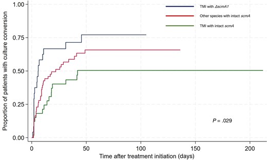
Kaplan-Meier curve for time to culture conversion. Abbreviation: TMI, typical Mycobacterium intracellulare.
Factors Associated With Culture Conversion
| Factor | Univariate Analysis | Multivariate Analysis | ||
|---|---|---|---|---|
| Unadjusted HR (95% CI) | P Value | Adjusted HR (95% CI) | P Value | |
| Age, y | 0.96 (.94–.99) | .004 | 0.97 (.94–.99) | .047 |
| Male vs female | 0.46 (.27–.78) | .004 | 0.53 (.30–.93) | .027 |
| BMI, kg/m2 | 1.10 (1.02–1.19) | .019 | 1.09 (.99–1.19) | .056 |
| AFB smear positivity | 0.69 (.41–1.19) | .186 | 0.98 (.54–1.77) | .940 |
| Presence of cavity | 0.40 (.26–.63) | <.001 | 0.41 (.25–.68) | .001 |
| Subspecies and S-CM truncation | ||||
| TIM with intact scmA | Reference | Reference | ||
| TIM with ΔscmA1 | 2.32 (1.18–4.56) | .015 | 2.17 (1.08–4.34) | .029 |
| Other subspecies with intact scmA | 1.52 (.85–2.74) | .155 | 1.21 (.66–2.22) | .531 |
Abbreviations: AFB, acid-fast bacilli; BMI, body mass index; CI, confidence interval; HR, hazard ratio; S-CM, secreted chorismate mutase; TMI, typical Mycobacterium intracellulare.
Apoptotic Cell Death in Murine Macrophages Infected With M intracellulare With or Without S-CM Truncation
Since S-CM promotes bacterial survival by inhibiting intrinsic macrophage apoptosis in M bovis BCG [9], we investigated whether the natural truncation of S-CM in TMI induces apoptosis and suppresses bacterial survival. Murine macrophage J774A.1 cells were infected with 2 TMI isolates possessing intact scmA and 2 isolates harboring the ΔscmA1 mutation. To exclude the possibility that differences in bacterial growth rates influenced infection outcomes, we first compared their growth curves and found no significant differences among the isolates (Supplementary Figure 3A).
At 24 hours postinfection, TMI strains with ΔscmA1 induced significantly higher proportions of early apoptotic cells (Annexin V+/7-AAD−) and late apoptotic cells (Annexin V+/7-AAD+) than those with intact scmA. By 48 hours postinfection, late apoptotic cell levels remained elevated in ΔscmA1-infected cells (Figure 2A). Consistent with these findings, intracellular bacterial burdens were consistently lower for ΔscmA1 strains at both 24 and 48 hours postinfection (Figure 2B). Moreover, extracellular CFU counts were also reduced in ΔscmA1 strains, indicating bacterial elimination rather than release from the cells (Supplementary Figure 3B). These results were reproducible in differentiated THP-1 cells, a human macrophage-like model, which also exhibited increased apoptosis and reduced bacterial burden following infection with ΔscmA1 strains (Supplementary Figures 3C and 3D).
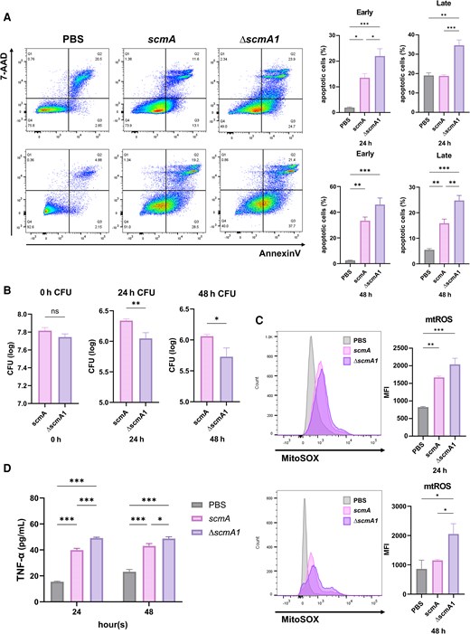
Apoptotic cell death in murine macrophages infected with Mycobacterium intracellulare strains harboring intact scmA or ΔscmA1. J774A.1 macrophage cells were infected with M intracellulare strains carrying intact or truncated secreted chorismate mutase at a multiplicity of infection of 10. A, Early and late apoptotic cells were assessed at 24 and 48 h postinfection using Annexin V/7-AAD staining. B, Intracellular bacterial burdens were quantified using colony-forming unit assays at 0, 24, and 48 h postinfection. C, Mitochondrial reactive oxygen species production was measured using MitoSOX staining at 24 and 48 h postinfection. D, Tumor necrosis factor alpha levels in culture supernatants were quantified at 24 and 48 h postinfection using an enzyme-linked immunosorbent assay. Data are presented as mean ± standard error of the mean from at least 3 independent experiments. Statistical significance was determined using 1-way analysis of variance with Tukey multiple comparisons test (A, C, D) or Student t test (B) (*P < .05, **P < .01, ***P < .001). Abbreviations: CFU, colony-forming units; MFI, mean fluorescence intensity; mtROS, mitochondrial reactive oxygen species; ns, not significant; PBS, phosphate-buffered saline; TNF-α, tumor necrosis factor alpha.
Expression of the anti-apoptotic gene Bcl-2 [21] was downregulated, whereas proapoptotic genes BAK and TNF-α [22] were upregulated in cells infected with ΔscmA1 strains (Supplementary Figure 3E). Consistent with our previous finding in M bovis [9], ΔscmA1 TMI strains induced significantly higher mtROS levels at 48 hours and markedly increased TNF-α secretion at 24 and 48 hours postinfection (Figures 2C and 2D). These effects were not observed with heat-killed strains, indicating that the observed responses depend on active bacterial processes rather than passive components (Supplementary Figure 3F).
Suppression of Apoptosis by scmA Complementation in ΔscmA1 TMI Strains
J774A.1 cells were infected with a recombinant M intracellulare ATCC13950T strain, a ΔscmA1 strain, harboring either an empty vector (pMV306 Ø) or a vector expressing scmA (pMV306_scmA) (Supplementary Figure 4A). At 24 hours postinfection, cells infected with the pMV306_scmA strain exhibited a statistically significant reduction in early apoptotic cells than those with the pMV306 Ø strain. By 48 hours postinfection, both early and late apoptotic cell levels were reduced (Figure 3A). The pMV306_scmA strain exhibited higher intracellular and extracellular bacterial burdens compared to those of the pMV306 Ø group (Figure 3B and Supplementary Figure 4B). Infections using the human monocytic THP-1 cell line yielded consistent results, with the pMV306_scmA strain showing both increased bacterial burden and reduced levels of apoptotic cells (Supplementary Figures 4C and 4D).
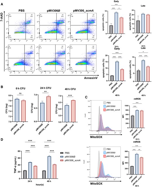
Secreted chorismate mutase expression suppresses apoptosis in Mycobacterium intracellulare ATCC 13950T, a ΔscmA1 strain. J774A.1 macrophage cells were infected with M intracellulare strains containing either the pMV306 or pMV306_scmA vector at a multiplicity of infection of 10. A, Early and late apoptotic cells were assessed at 24 and 48 h postinfection using Annexin V/7-AAD staining. B, Intracellular bacterial burdens were quantified using colony-forming unit assays at 0, 24, and 48 h postinfection. C, Mitochondrial reactive oxygen species production was measured using MitoSOX staining at 24 and 48 h postinfection. D, Tumor necrosis factor alpha levels in culture supernatants were quantified at 24 and 48 h postinfection using an enzyme-linked immunosorbent assay. Data are presented as the mean ± standard error of the mean from at least 3 independent experiments. Statistical significance was determined using 1-way analysis of variance with Tukey multiple comparisons test (A, C, D) or Student t test (B) (*P < .05, **P < .01, ***P < .00). Abbreviations: CFU, colony-forming units; MFI, mean fluorescence intensity; ns, not significant; PBS, phosphate-buffered saline; TNF-α, tumor necrosis factor alpha.
Similarly, cells infected with the pMV306_scmA strain showed significantly reduced mtROS levels at 48 hours and lower TNF-α expression at 24 and 48 hours (Figures 3C and 3D). Additionally, introduction of pMV306_scmA into clinical ΔscmA1 isolates suppressed apoptosis and restored bacterial survival, confirming the role of S-CM as an anti-apoptotic factor in M intracellulare (Supplementary Figures 5A and 5B).
DISCUSSION
This study shows that S-CM is a pathogenic factor in M intracellulare. Although scmA was consistently present across all analyzed M intracellulare strains, the ΔscmA1 mutant (C276A in the Rv1885c orthologue), which harbors a truncated form of S-CM, was exclusively observed in TMI strains. The presence of truncated S-CM significantly influenced clinical outcomes, with patients infected with ΔscmA1 mutant strains exhibiting milder initial disease severity, characterized by lower incidence of cavitation and improved early culture conversion rates. These findings indicate the pivotal role of S-CM in influencing disease severity and clinical outcomes in M intracellulare PD.
The pathogenicity of M intracellulare remains a debated topic, with reports showing variations in disease severity and treatment outcomes [5–7]. These inconsistencies likely arise from the existence of multiple subspecies within M intracellulare and differences in strain-specific pathogenicity [8, 23–25]. Furthermore, in addressing these variations, a comprehensive analysis of M intracellulare pathogenicity was conducted. In this study, we employed genotyping to classify M intracellulare subspecies and examined the diversity of scmA, a factor we hypothesized to influence pathogenicity [9, 10]. Our findings revealed that ΔscmA1 mutant occurred exclusively in TMI strains and was associated with differences in patient prognosis.
Approximately 20% of the isolates contained a truncated form of S-CM, resulting from a C276A mutation that introduces a premature stop codon. Strains with ΔscmA1 demonstrated significantly faster culture conversion rates. This observation is likely related to the greater disease severity associated with the presence of intact S-CM. Cavitation was more common in patients infected with TMI strains possessing intact scmA. Furthermore, these patients more frequently required medications such as clofazimine, which are typically reserved for severe cases [26], in patients infected with TMI strains, thereby supporting the connection between intact S-CM and increased disease severity. These findings suggest the role of intact S-CM in modulating disease severity and influencing treatment response.
The underlying mechanism by which S-CM influences disease severity and treatment response appears to involve the suppression of macrophage apoptosis, a process essential for their function in the innate immune response against mycobacteria [27]. Although apoptosis of macrophages is widely regarded as a defense mechanism against intracellular pathogens [28, 29], its role in NTM infections varies depending on the specific strain involved. For example, macrophage apoptosis has been shown to effectively suppress M avium by preventing the release of intracellular components and restricting bacterial spread, highlighting its protective roles [30]. In contrast, apoptosis promotes cell-to-cell spread of bacteria in M abscessus infection, thereby increasing its virulence [31]. In our study, the truncation of S-CM enhanced macrophage apoptosis, exhibiting a protective effect against M intracellulare.
To verify this, macrophages were infected with M intracellulare strains harboring either intact scmA or ΔscmA1 mutation. Strains with intact scmA suppressed the induction of mtROS and TNF-α, thereby reducing apoptotic cell death. In contrast, ΔscmA1 strains led to significant increases in both early and late apoptosis. These strains also downregulated the expression of Bcl-2 and upregulated BAK and TNF-α, indicating a proapoptotic and inflammatory phenotype. Notably, TNF-α induction was not observed in infections with heat-killed strains, confirming that the observed response depends on live bacterial activity.
To further investigate the role of S-CM, we introduced a scmA-expressing vector into ΔscmA1 strains, including the M intracellulare type strain and selected clinical isolates. Complementation with scmA reduced apoptosis and TNF-α levels while enhancing bacterial survival in both murine J774A.1 macrophages and THP-1–derived human macrophages. These findings align with our previous research, which shows that S-CM promotes bacterial survival by inhibiting macrophage apoptosis in M bovis BCG infection [9]. This conserved mechanism across different mycobacterial species highlights the significance of S-CM as a pathogenic factor in modulating immune response and influencing clinical outcomes.
In NTM-PD, disease severity, lung function, and treatment response are commonly predicted using clinical indicators such as age, sex, BMI, and radiologic findings [32–34]. Microbiologic factors remain poorly understood. We recently identified trehalose-dimycolate, a component of the mycobacterial cell wall, as a driver of proinflammatory responses and a favorable predictor of disease progression and treatment response in M avium complex PD [35]. Similarly, we propose that S-CM could serve as a microbiologic predictor of disease severity and treatment response in patients with M intracellulare PD, potentially enabling more personalized therapeutic strategies.
This study had some limitations. First, the role of S-CM in species other than M intracellulare was not evaluated, and the sample size was relatively small. Second, the inability to analyze isolates collected at the time of diagnosis prevented the assessment of the natural course of the disease before treatment. Third, the precise mechanism linking the enzymatic function of S-CM (catalyzing the conversion of chorismate to prephenate) to its role as a virulence factor that modulates apoptosis and promotes bacterial survival remains unclear. Finally, the differences between strains harboring intact scmA and those with ΔscmA1 were not examined in an in vivo infection model, which warrants future research. Despite these limitations, our integration of clinical data and experimental findings supports the role of S-CM as a novel microbiological factor in M intracellulare PD and highlights its potential as a prognostic biomarker in clinical practice.
In conclusion, the truncation of S-CM in M intracellulare enhances macrophage apoptosis, reducing bacterial burden. Consequently, strains with truncated S-CM exhibit lower pathogenicity and improved treatment responses. These findings suggest that S-CM may serve as a predictive marker for pathogenicity in future studies on M intracellulare PD.
Supplementary Data
Supplementary materials are available at The Journal of Infectious Diseases online (http://jid.oxfordjournals.org/). Supplementary materials consist of data provided by the author that are published to benefit the reader. The posted materials are not copyedited. The contents of all supplementary data are the sole responsibility of the authors. Questions or messages regarding errors should be addressed to the author.
Notes
Author contributions. N. K. and B.-J. K. conceived and designed the project. All authors contributed to the data acquisition. J. B., J.-Y. L., H. S., B.-J. K., and N. K. interpreted the data. All authors drafted and approved the manuscript.
Prior presentation. This study was presented as an abstract (number P2-17) at the 138th Congress of the Korean Academy of Tuberculosis and Respiratory Disease, Seoul, South Korea, 23–25 October 2024.
Data availability. Anonymized data can be shared upon appropriate request and with approval of the Institutional Review Board of Seoul National University Hospital. Data requests can be made to the corresponding authors.
Financial support. This work was supported by a grant of Patient-Centered Clinical Research Coordinating Center funded by the Ministry of Health and Welfare, South Korea (grant number RS-2021-KH119793 to J.-J. Y.). This work was also supported by a grant from the Korea Health Technology R&D Project through the Korea Health Industry Development Institute, funded by the Ministry of Health and Welfare, South Korea (grant number RS-2024-00456525 to B.-J. K.). This work was also supported by the “Korea National Institute of Health” (KNIH) research project (grant number 2024-ER2105-00 to N.K). [KNIH is the branched organization of K-CDC].
References
Author notes
J. B., J.-Y. L., and H. S. contributed equally to this work.
Potential conflicts of interest. J.-J. Y. has participated as the overall or institutional principal investigator in multiple clinical trials sponsored by Insmed, AN2 Therapeutics, and LigaChem Biosciences. All other authors report no potential conflicts.
All authors have submitted the ICMJE Form for Disclosure of Potential Conflicts of Interest. Conflicts that the editors consider relevant to the content of the manuscript have been disclosed.


