Cover image

Diagnosis of malignant coronary vasospasm by 12-lead Holter electrocardiogram and optical coherence tomography
Sarah Bär1†, Tilman Perrin2†, Lorenz Räber1, and Tobias Reichlin1*
1Department of Cardiology, Swiss Cardiovascular Center, Inselspital, University Hospital Bern, University of Bern, 3010 Bern, Switzerland; and 2Department of Cardiology, Bürgerspital Solothurn, 4500 Solothurn, Switzerland
* Corresponding author. Tel: 141 31 664 00 50, Email: [email protected]
†Sarah Bär and Tilman Perrin authors contributed equally to this article.
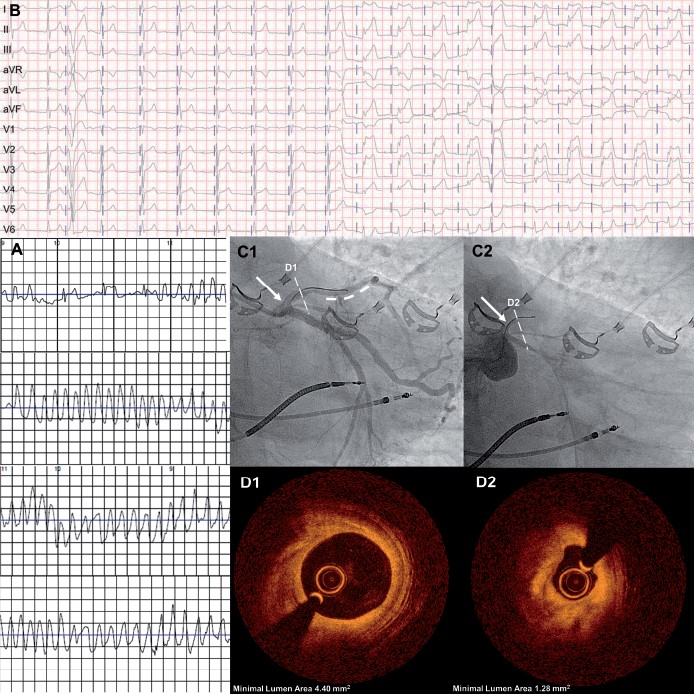
A 73-year-old man presented with recurrent syncopes preceded by angina. Three years earlier, the patient had a pacemaker implanted due to symptomatic sick sinus syndrome. Device interrogation showed several torsade-like ventricular tachycardias (VTs) corresponding with the symptoms. Left ventricular ejection fraction was normal. Coronary angiography revealed diffuse coronary sclerosis and a hazy 50% lesion in the mid left anterior descending artery (LAD), which was treated with a drug-eluting stent. Three days later, the patient experienced another syncope with documentation of ventricular fibrillation by the provided life-vest (Panel A). The pacemaker was upgraded to an implantable cardioverter-defibrillator. During subsequent workup, a 12-lead Holter electrocardiogram (ECG) revealed intermittent giant ST-segment elevations in the anterior and inferior leads (Panel B) with concurrent angina. Repeat coronary angiography showed a good result of the LAD stent (Panel C1, bold dashed line) and normal minimal lumen area of LAD by optical coherence tomography (OCT) (Panel D1). Intracoronary application of 50 µg methergin triggered subtotal left main coronary artery vasospasm (Panel C2) with global ST-segment elevations, severe hypotension and non-sustained VT. The beginning of the spasm was documented by OCT showing a thickening of the media with consecutive lumen narrowing (Panel D2). The vasospasm was reverted by application of sublingual isosorbiddinitrat and intra-coronary nitroglycerine. Repeat testing thereafter was uneventful. A treatment with amlodipine and molsidomine was introduced before discharge. The patient remained symptom-free for 3 months and a 12-lead Holter ECG showed no further ST-segment changes. According to our case, both 12-lead Holter ECG and coronary vasospasm testing should be considered during workup of patients with angina without significant coronary artery disease.


