


 https://orcid.org/0000-0003-3160-9022

This scientific commentary refers to ‘Imaging subtypes reveal distinct biological substrates and disability profiles in multiple sclerosis’, by Preziosa et al. (https://doi.org/10.1093/brain/awag320).
Multiple sclerosis (MS) is highly heterogeneous. Patients differ in clinical manifestations, inflammatory activity, treatment response and the relative contributions of focal lesions, diffuse white matter injury and grey matter neurodegeneration. Clinical phenotypes, particularly relapsing-remitting and progressive forms, remain useful but do not fully capture this biological diversity. The coexistence of inflammatory and neurodegenerative processes throughout the disease course has prompted calls for a more mechanism-driven classification.1 Patients with similar clinical labels may differ substantially in the processes contributing to disability, and suppressing focal inflammatory activity may leave other sources of tissue injury insufficiently addressed. As disease-modifying therapies become more effective, a biologically informed classification could help refine disability risk estimates and guide treatment by clarifying the processes driving disease in individual patients. Such classification could also improve patient selection and stratification in clinical trials, define more homogeneous groups for biomarker and genetic studies, and support re-evaluation of previous findings in light of disease heterogeneity.
In this issue of Brain, Preziosa and colleagues2 advance this effort using multimodal MRI and Subtype and Stage Inference (SuStaIn). Unlike conventional clustering, SuStaIn reconstructs alternative trajectories of abnormality accumulation and estimates how far an individual has progressed along the most likely trajectory.3 In other words, the method aims to determine whether differences between patients reflect more advanced disease, a different sequence of tissue damage, or both. The authors analysed 3 T MRI scans from 1017 patients with MS and 548 healthy controls, integrating T2 lesion volume, mean diffusivity within lesions, normal-appearing white matter and white matter tracts, and regional brain volumes. Longitudinal clinical data, with median follow-up exceeding 6 years, were available for a subset of participants. Four MRI-defined subtypes emerged: lesion-led (44%), cortex-led (23%), tract-led (23%) and deep grey matter/cerebellar-led (10%). The model suggested that the earliest abnormalities were lesion accumulation in the lesion-led subtype, cortical atrophy in the cortex-led subtype, diffuse white matter microstructural abnormalities in the tract-led subtype, and thalamic, hippocampal and cerebellar atrophy in the deep grey matter/cerebellar-led subtype. Application of the model without retraining identified all four subtypes in an independent cohort of 247 patients and 141 healthy controls.2
The subtypes differed in their clinical profiles. Progressive MS was more common and disability was greater in cortex-led and deep grey matter/cerebellar-led disease, although the association with progressive disease weakened after adjustment for demographic factors. Tract-led disease was enriched in paediatric-onset MS, whereas deep grey matter/cerebellar-led disease was relatively more common in late-onset MS. Progression independent of relapse activity (PIRA) occurred earlier in cortex-led and deep grey matter/cerebellar-led disease than in lesion-led disease, although these differences also decreased after adjustment for age and sex. Cognitive performance did not differ significantly between subtypes; however, higher SuStaIn stage was associated with poorer cognitive performance.2 Together, these findings suggest that the subtype and stage of brain tissue alterations are associated with different clinical characteristics.
How should these four patterns be reconciled with earlier classifications (Fig. 1)? Eshaghi and colleagues4 identified cortex-led, normal-appearing white matter-led and lesion-led subtypes using T2 lesion volume, regional grey matter volumes and regional normal-appearing white matter T1/T2 ratios. Pontillo and colleagues,5 using lesion burden and regional grey matter volumes, identified deep grey matter-first and cortex-first subtypes. The tract-led subtype identified by Preziosa and colleagues2 resembles the previously described normal-appearing white matter-led pattern, whereas the deep grey matter/cerebellar-led subtype may distinguish a more specific anatomical pattern within earlier grey matter-based classifications. These similarities suggest some overlap between classification systems. However, similarly named subtypes may represent different patient groups, and identifying more subtypes does not necessarily imply greater biological validity.
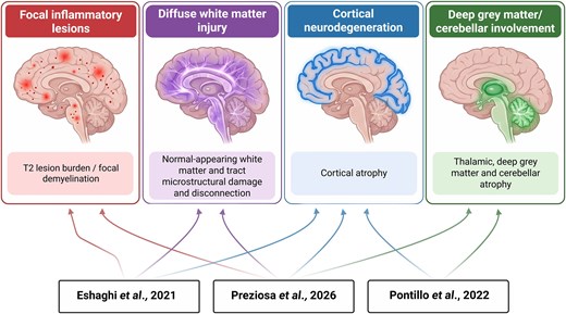
Biological heterogeneity of multiple sclerosis and MRI-derived SuStaIn subtypes. Four overlapping dimensions of tissue damage in multiple sclerosis are schematically illustrated. Colours indicate conceptual similarities across studies,2,4,5 without implying equivalent patient groups or mutually exclusive pathological processes. Differences between classifications may reflect imaging inputs, cohort characteristics and modelling choices. Subtype terminology has been harmonized for comparison: the cortex-first and deep grey matter-first subtypes of Pontillo et al.5 are shown as cortex-led and deep grey matter-led, respectively. Created using ChatGPT (OpenAI) and BioRender. Uher, T. (2026) https://BioRender.com/r7pk4si.
SuStaIn compares candidate models with different numbers of trajectories, using cross-validation to assess how well they fit the data and favouring simpler models when additional complexity is poorly supported.3 Applying the SuStaIn model to an independent cohort without retraining is therefore an important strength of the present study.2 However, the number of subtypes identified may still depend on sample size, population heterogeneity, the biomarkers included, and the reference populations and thresholds used to define abnormality. Future studies could build on this work through sensitivity analyses examining whether subtype number and assignment remain stable across reasonable changes in these choices. Independently fitting models in other cohorts would further assess whether similar subtypes and sequences emerge, while reporting assignment probabilities would indicate how confidently individual patients fit each subtype.
Including additional biological measures can also change the classification. Willard and colleagues6 combined MRI with serum neurofilament light chain concentrations and identified patterns with early or late biomarker elevation and different sequences of white and grey matter injury. Adding serum neurofilament light chain improved correlations between stage and disability, and the resulting subtypes differed in subsequent radiological activity.6 These findings suggest that SuStaIn subtypes depend partly on the measurements included in the model, and further evidence is needed to determine whether they reflect distinct disease mechanisms.
Assessing clinical usefulness also requires distinguishing prognosis from prediction of treatment benefit. Jiang and colleagues7 reproduced differences in MRI severity and inflammatory activity across the original MRI-defined subtypes. However, the subtypes did not convincingly predict confirmed disability progression, and treatment effects did not differ significantly between them.7 A treatment effect reaching statistical significance in one subgroup but not another does not establish that the effects differ.8 Demonstrating differential benefit requires an adequately powered Treatment × Subtype interaction test, preferably prespecified.9 Conversely, a non-significant interaction does not establish equivalence, particularly when subgroup sizes or event counts are small.
Disease severity and the rate of disease progression will affect the ability to detect clinically meaningful differences between subtypes. Interpretation of prognostic differences must also consider treatment exposure, which may modify the outcomes associated with each subtype. For example, earlier work associated the lesion-led subtype with greater inflammatory activity and less favourable outcomes,4 whereas patients assigned to this subtype in the present cohort had lower disability and longer PIRA-free survival than those assigned to the cortex-led or deep grey matter/cerebellar-led subtypes.2 Differences in treatment exposure and the extent to which inflammatory activity was suppressed could partly explain this contrast, alongside differences in cohort characteristics and outcome definitions. However, these comparisons cannot establish whether treatment affects disease trajectories differently across subtypes. The prognostic value of an imaging subtype may therefore depend partly on the treatment received.
The model’s assumptions deserve equal attention. SuStaIn allows different sequences between subtypes but assumes a shared underlying sequence within each subtype.3 Biomarker abnormalities are assumed to remain stable or progress monotonically with model stage, without requiring a constant rate of change. However, this assumption does not capture biological improvement, such as partial resolution of T2 lesions. Apparent improvement, such as an increase in measured brain volume, may instead reflect measurement error. Moreover, differences between individuals observed cross-sectionally need not reproduce changes within an individual over time.10 Longitudinal validation, as undertaken in earlier SuStaIn studies,4 helps address this concern but does not establish lifelong adherence to an inferred trajectory. The term ‘led’ denotes the earliest abnormalities in the modelled sequence, not necessarily the tissue showing the fastest current deterioration. A tract-led patient could therefore develop substantial cortical atrophy without contradicting the original classification. The key questions are whether subtype assignments remain reproducible on repeated scans and whether subsequent changes follow the predicted sequence, while accounting for uncertainty in both stage and subtype.
Addressing these questions will require standardized longitudinal MRI beginning early in the disease course, linked to detailed treatment histories and clinical outcomes, with analyses that account for potential confounders. Spinal cord imaging, fluid biomarkers and measures of cortical or chronic active lesions could capture important aspects of disease not reflected by brain volumetry and conventional lesion assessment, although their inclusion should be justified by reproducibility and added clinical value. Genetic and other biological comparisons across MRI subtypes may help clarify whether different anatomical patterns reflect distinct disease mechanisms. To establish added prognostic value, prospective studies should compare subtype-informed models with models based on age, disease duration, disability and conventional MRI measures, and assess both how closely predicted risks match observed outcomes and how well the models distinguish patients at higher and lower risk. For treatment selection, the decisive test is whether incorporating subtype improves clinical decisions and patient outcomes. Validation in independent populations should assess potential overfitting and establish whether these benefits persist across centres, scanners and treatment eras.
Preziosa and colleagues2 have identified four patterns of brain damage in MS that may reflect different sequences of tissue injury. Their findings support moving beyond conventional clinical phenotypes, while highlighting the evidence still needed before MRI-based subtyping can be used clinically. With further validation, a biologically informed MRI classification could help link clinical course to underlying disease mechanisms and guide treatment selection for individual patients.
Funding
T.U. is funded by the Ministry of Health of the Czech Republic, the institutional support of a hospital research project (MH CZ-DRO-VFN64165), the Charles University Cooperation Program in Neuroscience and the Czech Ministry of Health project (NU22-04-00193). G.N. is funded by a senior clinical investigator fellowship of Fonds Wetenschappelijk Onderzoek / Research Foundation Flanders (1805620N, www.fwo.be).
Competing interests
G. N. was a minority shareholder in icometrix until 2025.


