


Abstract
Leishmaniasis is a disease predominantly prevalent in the tropics, considered as one of the primary neglected diseases, preferably affects individuals of low socioeconomic status. Although this condition is well described in children, disseminated cutaneous leishmaniasis is a rare form of increasing importance and multiple cases observed in the adult population; however, still little described in children.
We present the case of a 12-year-old male, who has multiple ulcerative and nodular lesions distributed throughout the body, of ∼1 year of evolution that did not respond to antimicrobial treatment. After the diagnostic process, positive serological tests were found for leishmaniasis, with improvement in the picture after the use of sodium stibogluconate.
Disseminated cutaneous leishmaniasis is a clinical form that is described with increasing frequency and should be recognized and treated appropriately, mainly in the pediatric population, avoiding complications and sequelae.
INTRODUCTION
Leishmaniasis is a globally distributed neglected disease that is caused by ∼20 species of the genus Leishmania distributed worldwide [1], which are transmitted through vectors by at least 30 species of phlebotomine, Lutzomyia in America and Phlebotomus in the rest of the world [2].
Leishmaniasis is one of the seven most critical neglected diseases, preferably affecting individuals of low socioeconomic status; it is widely distributed in about 88 countries, preferably in tropical and subtropical climates, with >350 million people at risk of contracting the disease [3]. In Peru, American Tegumentary Leishmaniasis (ATL) is considered the second most prevalent tropical disease; the first is dengue. Also, it is the third most common infectious disease in terms of morbidity, after tuberculosis and malaria. There are eight disease-causing species with varied geographical distribution and clinical characteristics [4].
It is known that different species of Leishmania can cause various clinical manifestations, and the severity varies from spontaneously healing lesions to life-threatening visceral disease. The outcome is determined by the inter-relationship between the characteristics of the parasite, the biology of the vector and the immune response of host [1].
Disseminated cutaneous leishmaniasis (DCL) is a rare clinical spectrum of leishmaniasis, with <2% of total cases affected with ATL and is characterized by the appearance of dozens to thousands of polymorphic lesions in various parts of the body [5, 6]. This variant has been very little reported in children, so we present the case of a pediatric patient with a diagnosis of DCL.
CASE
Male patient, 12 years old, originally from and resident in the city of Huánuco, located in the central region of Peru at 1918 m above sea level. He made periodic trips to the town of Aucayacu, a tropical jungle region 113 km away. He went to the outpatient infectious disease service of the Hospital II EsSalud (Social Security Health), in Huanuco, because he presented multiple ulcerous and nodular skin lesions with regular edges and over elevated, distributed in the anterior and internal region of the right thigh, left thigh, gluteal level, in the left forearm, back and abdomen, being the largest located in the anterior face of the right leg of 3 cm × 2 cm and the smallest in the paravertebral region of 1 cm × 1 cm (Fig. 1). His relative indicates that some of them presented seropurulent secretion. At the physical examination, the lesions were not painful on palpation, with no signs of phlogosis or secretion. The initial wound appeared on the right thigh ∼1 year earlier; then, gradually appeared on various parts of the body. Between December 2018 and August 2019, he received topical antibiotics and oral clindamycin for ten days in up to three therapeutic courses, with no response or improvement.
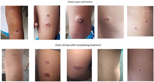
Evolution of lesions with sodium stibogluconate treatment.
The patient denied any history of significance, did not use chronic medication, and at the time of the physical examination on admission had no oral or nasal mucosal lesions and no history of previous respiratory discomfort.
An indirect immunofluorescence (IFA) method was performed; this was positive (1/40), so a biopsy of one of the lesions is performed, and treatment with sodium stibogluconate was initiated at a dose of 20 mg/kg/day intravenously for 30 days. The family member refers that 10 days after the beginning of the treatment, the patient presents headache, vomiting and choluria. He is admitted to our hospital on 1 August 2019, as an emergency patient, where he was given the treatment of hydration, antiemetics and analgesics with overall improvement and was discharged the following day. Treatment with stibogluconate was not suspended.
Biopsy results reported ulcerated skin to hypodermis with granuloma formation with multinucleated Langhans type giant cells, without observation of parasites. No molecular tests or tissue culture were performed because this technology was not available at our hospital. Tests for HIV, HTLV, Hepatitis B and Hepatitis C infection were negative, as well as the chest X-ray and three sputum smears for tuberculosis. His hepatic and renal tests were normal during the treatment and follow-up.
At the end of treatment, smooth, depressed and hypochromic scars were observed on the forearm, abdomen and back. Injuries to the right thigh showed slower healing than other wounds. One month after treatment was completed, there was a significant improvement in the leg lesions with complete healing of the back, arm and abdominal lesions.
DISCUSSION
In Peru, leishmaniasis is one of the most relevant endemic diseases. Peru is considered one of the ten countries with more cases in the world [2]. Clinically, it presents two very characteristic and differentiated forms: the Andean cutaneous leishmaniasis or ‘Uta’, and mucocutaneous leishmaniasis [4].
DCL is an emerging form of ATL, defined by the presence of more than ten acneiform, papular and ulcerated lesions on at least two parts of the body. In most cases, DCL initially presents as an ulcer-type lesion with the subsequent appearance of other polymorphic lesions in different places to the initial lesion [7]. However, this form has been little documented or reported in our country [8].
It has been suggested that the forms of DCL have increased due to the presence of different degrees of immunosuppression and even malnutrition [9]. However, there have been some studies that showed that the parasites involved in cases of DCL, induce different responses in neutrophils of healthy patients, suggesting that certain parasite species determine the final clinical presentation [7]. Also, multiple clones of Leishmania braziliensis species are involved in the pathogenesis of different clinical forms of leishmaniasis, and cases are frequently described in immunocompetent patients [5]. In our region, studies have been published showing that the species with the highest isolation is L. braziliensiş and even clinical hybrid forms of coinfecting species, L. braziliensis and Leishmania peruviana [10]. The only case of DCL previously reported in our country corresponded to a 62-year-old male in Ancash that tested positive for L. peruviana [8].
We found that the biopsy was unable to demonstrate the parasite, similar to other reports of DCL [11]. A recent review of the disease indicated that it is remarkable that in patients with DCL, it is not easy to find parasites in the skin samples [5]. A Peruvian study found that 27% of all patients who had tested negative to the direct microscopic test were under 19 years old [12]. It is also remarkable that other authors have also found multinucleated giant cells and granulomas in the respective biopsies [13, 14], and one of them refers to that in 43% of the biopsies granulomas were observed without observation of the parasite [15].
Indirect IFA is a serological technique of suitable sensitivity and specificity for leishmaniasis [16]. Like us, other studies initiated the diagnostic and therapeutic management after the results of this test, suggesting that the IFA technique is a useful method for diagnosis, control and clinical cure monitoring [17]. Nevertheless, some reports indicate that it can yield some false-negative results [11].
Described cases of DCL in the pediatric population are even rarer. An important feature is that, as with our patient, adverse effects to treatment with stibogluconate are relatively frequent in children [18], then the administration of this drug should be carefully monitored in the pediatric population.
Notably, ∼50% of the cases showed little response to initial treatment with antimonials, requiring the use of amphotericin, miltefosine and even fluconazole and ketoconazole [13, 19–21]. These findings suggest that in children with DCL, an adequate response to the use of antimonials should be promptly assessed, and the use of alternative medications also considered.
On the contrary, in most of the described cases of DCL of the adult population, there is an excellent response to the treatment with antimonials, being even one of the ways to differentiate it from the anergic diffuse cutaneous leishmaniasis [19, 22]. In the case of our patient, he presented adequate healing 30 days after completing treatment with sodium stibogluconate, which would be explained in part due to his age, almost a teenager, compared with other younger cases with less clinical response.
Another issue that must be assessed and that, unfortunately, is not commonly performed, is the identification of the species. Of the four studies that include seven pediatric patients, the species were only confirmed in three of them. Two patients from America were L. brazilensis, and in one case reported in Iran, Leishmania major [13, 19–21] was isolated.
When compared with the situation in adults, leishmaniases are neglected in children and adolescents [23], and in countries such as Peru, are still highly prevalent, with atypical forms, such as DCL, making challenging the diagnosis and treatment. We conclude that DCL is a clinical form that is more frequently seen in endemic areas. Although it is rarely described in the pediatric population, it should be recognized early and treated appropriately to avoid complications and sequelae in this type of patient.

Comments