


A 69-year-old woman with no significant medical history presented to our department with bilateral scleritis, posterior auricular pain and bilateral jaw pain 8 months earlier. Her serum C-reactive protein level was 2.55 mg/dl. Her erythrocyte sedimentation rate was 65 mm/h, and anti-nuclear antibody, rheumatoid factor and anti-neutrophil cytoplasmic antibodies were negative. Ultrasonography showed wall thickening of the left posterior auricular artery (Fig. 1A) and tenosynovitis of the long head of both biceps (data not shown). T2-weighted magnetic resonance imaging indicated wall thickening in the left posterior auricular artery (Fig. 1B). Increased 18F-fluorodeoxyglucose uptake in the bilateral walls of the brachiocephalic arteries and shoulder joints was observed with positron emission tomography combined with CT (data not shown). The patient was diagnosed with giant cell arteritis (GCA), and her symptoms rapidly improved with oral prednisolone (50 mg per day).
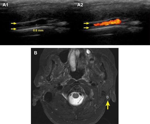
Wall thickening of the left posterior auricular artery
(A) Ultrasonography of the left posterior auricular artery in longitudinal view in B-mode (A1) and with colour Doppler (A2) showed wall thickening (0.6 mm, arrow). (B) T2-weighted magnetic resonance imaging indicated wall thickening in the left posterior auricular artery (arrow).
To the best of our knowledge, there have been no reports of GCA involving the posterior auricular artery [1, 2]. This artery branches from the external carotid artery and anastomoses with branches of the superficial temporal artery. GCA can affect the posterior auricular artery, and posterior auricular pain may be a clinical sign for the early diagnosis of GCA.
Acknowledgements
We gratefully acknowledge the work of past and present members of our department.
Funding: No specific funding was received from any bodies in the public, commercial or not-for-profit sectors to carry out the work described in this article.
Disclosure statement: The authors have declared no conflicts of interest.

Comments