


Background: Giant cell arteritis (GCA) affects both the cranial and large vessels. Ultrasonographic studies have reported that the incidence of large vessel vasculitis (LVV) in patients with GCA varies from 30% to 55%. The aim of this study was to investigate the rate of LVV in patients with GCA by using the new anteromedial approach to examine the large supraaortic vessels in addition to the cranial arteries.
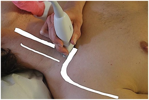
Methods: Patients with new-onset GCA referred to Department of Rheumatology, Martina Hansens Hospital in Bærum, Norway between September 2017 and November 2018 were examined. The diagnosis was based on the typical clinical manifestations, ultrasound findings and increased C-Reactive protein (CRP). All the patients were scanned by ultrasound, using the anteromedial approach for the supraaortic vessels (carotid, vertebral, subclavian, axillary proximally and distally), in addition to the examination of the cranial vessels (temporal, facial, occipital). The anteromedial approach consists of a continuous ultrasound evaluation of the large supraaortic vessels with the patient in a prone position (fig. 1). The examination utilized a GE S8 ultrasound machine with a 9-12 Mhz linear probe for the large vessels and a 18Mhz hockey stick probe for the cranial arteries. The age, gender, CRP and the distribution of vasculitis in the vessels were recorded.
Results: Twenty-eight patients, 24 (85%) females and 4 (25%) males, were diagnosed with GCA during the recruitment period. The mean age was 72 years (95% CI (68-76)). Mean CRP was 74 mg/dl (95% CI (54-93)). Of the 28 GCA patients, 6 patients (21%) had cranial GCA only, 7 patients (25%) had LVV only and 15 patients (54 %) had both cranial and LVV GCA. The temporal arteries were the most frequently affected in 57.1 % of the patients, followed by the facial (left 32, 1% and right 25%) and occipital (left 21.4 % and right 14.3%). The large supraaortic vessels mostly affected were the right proximal axillary (39.3%), the left distal axillary (39.3 %), the subclavian (left 35.7% and right 35.7 %), the left proximal axillary (35.7%) and the right distal axillary (35.7 %). The left carotid and vertebral was affected in 32.1 % of patients, while the right carotid in 3.6% and right vertebral in 17.9% of the patients.
Conclusion: The anteromedial ultrasound examination revealed inflammation of the large supraaortic vessels in 79% of the GCA patients. This indicates that the involvement of supraaortic arteries is more common in GCA than previously reported.
Disclosures: Andreas Diamantopoulos Speaker Honoraria Roche

Comments