


Individuals with systemic sclerosis often exhibit salt-and-pepper pigmentation, with a prevalence ranging from 51% to 94% in darker phenotypes.1 Interestingly, normal pigment preservation over superficial veins within these areas of altered pigmentation is described, although uncommon.
Case 1: A 43-year-old female, presented with Raynaud’s phenomenon, thickening and tightening of skin on her face and limbs; and digital tip ulceration with pitted scars for 7 years. Clinical examination revealed widespread sclerosis, salt-and-pepper pigmentation on various body parts, mask-like facies and sclerodactyly. Both legs exhibited areas of spotty depigmentation, and linear hyperpigmented streaks along superficial veins (Figure 1a).
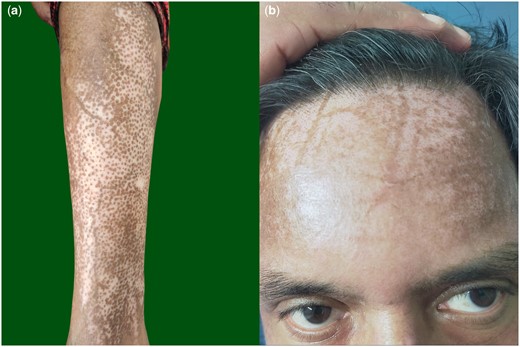
(a) Salt-and-pepper pigmentation over legs with sparing of pigmentation along superficial veins. (b) Pigment sparing over the course of superficial veins within areas of salt-and-pepper pigmentation on the forehead and scalp.
Case 2: A 45-year-old male presented with progressive tightening of the skin on his face, trunk for 9 months. Over the forehead and legs, we observed salt-and-pepper pigmentation. Along the course of his superficial veins on both forehead and frontal scalp, notably, there was a distinct preservation in pigmentation (Figure 1b).
In 1984, researchers first noticed pigment retention along superficial blood vessels within a larger field of depigmentation in three patients with systemic sclerosis; they also confirmed a simultaneous temperature difference between the pigmented and depigmented areas in one of these individuals.2 Plausibly, localized hyperthermia induces a specific reduction in the production of melanogenesis-inhibiting cytokines; leading to localized hyperpigmentation.
Researchers have rarely observed pigment retention over superficial veins in various other skin conditions, including vitiligo, dermatitides, dermatophytic infections, systemic lupus erythematosus and Rheumatoid arthritis.3,4 They hypothesize that a significant inflammatory response could induce vasodilation which would increase vascular permeability and activate endothelial cells. This leads to endothelial injury coupled with subclinical-to-mild phlebitis; the outcome is basal epidermal hyperpigmentation along with dermal pigment incontinence.4 Certain chemotherapy agents, including 5-fluorouracil, docetaxel, cyclophosphamide, bleomycin and vinca alkaloids; provoke serpentine supravenous hyperpigmentation after intravenous administration.3
In conclusion, the systemic sclerosis cases highlight the distinctive interplay of salt-and-pepper pigmentation, notably sparing pigmentation over superficial veins. Further exploration of the molecular and physiological aspects of these findings could contribute to a deeper understanding of the pathogenesis of pigmentary changes in systemic sclerosis.
Author contributions
Manavi Gupta (Conceptualization [equal], Writing—original draft [lead], Writing—review & editing [lead]), Shikha Shah (Conceptualization [lead], Data curation [lead]), and Tarun Narang (Supervision [lead], Validation [lead])
Funding
None declared.
Conflict of interest
None declared.
