


A 74-year-old male presented with a month-long history of a widespread erythematous rash and itchy brownish papules, previously diagnosed as pemphigus. Oral prednisone brought no improvement.
Physical examination revealed plaques of varying sizes and numerous quantities, with unevenly distributed black-brown papules and patches on the face, limbs and trunk, surrounded by erythematous areas. The surface of the patches is rough and exhibits wart-like growth, resembling rupioides proliferation (Figure 1 & 2).
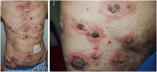
Lesions on the trunk and thighs, manifested as unevenly sized erythematous patches, with rupioides papules visible at the center of the patches.
Blood tests, including routine ones, blood glucose and liver and kidney function, were normal. The squamous cell carcinoma antigen showed a slight elevation (2.05 ng/ml), while pemphigus and pemphigoid antibodies were within normal limits. Imaging studies, including CT scans and MRI, found no malignancy evidence. Skin histopathology showed mild epidermal hyperplasia with keratinization, mucin infiltration and pseudo-horn cysts. The dermis exhibited abundant inflammatory cell infiltration, and direct immunofluorescence was negative.
The patient was diagnosed with Leser–Trélat sign, characterized by a rapid increase or growth of seborrheic keratosis accompanied by itching. Literature reports that skin manifestations resembling seborrheic keratosis clinically can be confused with pemphigus, especially in elderly patients.1 In our reported case, inflammatory erythema developed around the seborrheic keratosis, leading to a rash resembling crusty proliferative pemphigus. However, pathological examination and bridge granule protein antibody testing ruled out pemphigus lesions. Furthermore, Leser–Trélat sign is considered a paraneoplastic rash with a higher association with malignant tumors, particularly adenocarcinomas, although some patients may not have an associated malignancy.2 Therefore, comprehensive skin biopsy and anti-desmoglein antibody testing should be conducted in addition to systematic examinations to exclude malignancy in elderly individuals with a diffuse seborrheic keratosis-like rash.
Author contributions
Wenting Hu (Conceptualization [equal]) and Yangyang Ma (Project administration [equal])
Conflict of interest
None declared.
