


Japanese spotted fever (JSF) is an emerging rickettsiosis in Japan. Similar to other rickettsiosis, JSF is characterized by a triad of high fever, rash and eschar formation. Although pulmonary involvement and radiographic abnormalities are a well-documented complication of other rickettsiosis, limited cases of pulmonary involvement of JSF have been reported. In endemic areas of rickettsia, when the patients with pulmonary symptoms and radiographic findings of unknown cause are present, clinicians should consider rickettsial infection as a possible differential diagnosis.
Case presentation
A 75-year-old man with a 5-day history of fever, rash, cough and progressive dyspnea presented to the emergency department. He was diagnosed as having chronic obstructive pulmonary disease (COPD), but he had no previous cardiovascular, gastrointestinal problems or allergy. His temperature was 38.9°C, blood pressure was 109/74 mmHg, respiratory rate was 19 breaths/min and pulse was 94/min. On examination, the patient had a faint maculopapular rash on the trunk and extremities (Figure 1A). He was a farmer and lived in near wooded areas where ticks might have been present, but did not remember a tick bite. Laboratory examination showed thrombocytopenia, elevated liver enzyme levels, bilirubin and fibrinogen degradation product levels, and prolonged activated partial thromboplastin time and prothrombin time. Under the tentative diagnosis of rickettsiosis, scrub typhus or Japanese spotted fever (JSF), intravenous minocycline was started. Chest computed tomography (CT) revealed bilateral interstitial infiltrate, ground–glass opacities, interlobular septal thickening and consolidation (Figure 1B). Although, polymerase chain reaction for the serum sample was positive for Rickettsia japonica. His additional laboratory examination showed unremarkable changes including eosinophil count, KL-6 and serum IgE level. Based on the clinical and radiographic findings, he was diagnosed of JSF with pulmonary involvement (rickettsial pneumonia). His symptoms were gradually improved and subsequent CT findings faded without remission (Figure 1C).
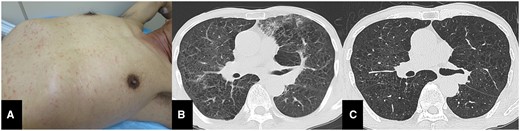
(A) Photographs of the patient’s trunk with maculopapular rash. (B) Chest CT showed bilateral interstitial infiltrate, ground glass opacities, interlobular septal thickening and consolidation. (C) Repeat chest CT showed that radiographic abnormalities were vanished after the treatment.
Discussion
JSF was discovered as an emerging rickettsiosis in Japan in 1984.1 The annual number of cases of JSF has been increasing three-fold compared to decades ago.2 Similar to other rickettsiosis, JSF is characterized by a triad of high fever, rash without itching and eschar formation. Most patients do not exhibit the classic features of JSF on their first visit to a medical institution. Other symptoms of JSF are often vague and nonspecific, including headache, fever, nausea, vomiting, myalgia and chills. Spotted fever group rickettsiosis generally invades various eukaryotic host cells, such as reticuloendothelial cells and vascular endothelial cells, and can spread the whole body.3 The infection of these cells increases vascular permeability of the blood vessels, which can cause several symptoms, such as fever, myalgia, nausea and skin rashes, and affect many organs. Pulmonary involvement and radiographic abnormalities are a well-documented complication of Rocky Mountain spotted fever, scrub typhus, Lyme disease and Babesiosis.4,5 However, limited cases of pulmonary involvement of JSF have been reported. Rickettsial pneumonia can be misdiagnosed as other atypical pneumonia or intestinal pneumonia due to radiographic findings and pulmonary symptoms. These findings are associated with non-cardiogenic pulmonary edema consequent to capillary endothelial damage due to rickettsial infection. β-lactam antibiotics and aminoglycosides, commonly used in the empirical treatment for febrile disease, are completely ineffective for rickettsial infection. Diagnostic delay sometimes leads to serious complication and death. Therefore, when the patients in endemic areas with pulmonary symptoms and radiographic findings of unknown cause are present, clinicians should consider rickettsial infection as a possible differential diagnosis.
Conflict of interest. None declared.
References
National Institute of Infectious Diseases (JP). NESID Annual Surveillance Data (Notifiable Diseases) 2014-2 [Internet]. http://www.nih.go.jp/niid/en/survei/2085-idwr/ydata/6058-report-ea2014-20.html (27 November 2020, date last accessed).
