


Ecthyma gangrenosum (EG) is a skin manifestation that initially appears as macular, erythematous lesions that becomes haemorrhagic and transforms into tender erythematous areas with necrotic centres.1 EG lesions often present on the extremities. EG is typically associated with Pseudomonas aeruginosa.2 However, these lesions can be seen with various Gram-positive and Gram-negative bacteria, and fungi such as Fusarium species.3 Infectious organisms enter via direct inoculation of the skin or through haematogeneous seeding to the affected site. They invade the local vasculature, causing a vasculitis and necrosis of local tissue. Particularly with immunosuppression, this can lead to disseminated infection via ongoing perivascular invasion. In immunocompromised patients, infection typically begins with direct inhalation or inoculation with the Fusarium conidia, which germinate and invade tissue.4 Fevers and cutaneous skin lesions are common primary manifestations.5 The central necrosis is suggestive of angioinvasion by the hyphae with associated thrombosis.5
The above patient was a 73-year-old man with chronic lymphocytic leukaemia and metastatic melanoma on nivolumab who presented to the hospital with neutropenic fevers. The patient developed a lesion on the left dorsal foot, appearing as a flat one-centimetre necrotic area with surrounding erythema (figure 1A). Blood cultures were obtained. A punch biopsy was also obtained and sent for pathological and microbiological studies. Grocott-Gomori's methenamine silver stain at ×64 magnification revealed acute angle-branching septated hyphae with angioinvasive components (figure 1B). A lactophenol cotton blue preparation at 40x-magnification demonstrated multiple macroconidia and microconidia (figure 1C), consistent with Fusarium. Interestingly, all cultures obtained were unable to yield any bacterial growth.
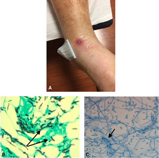
The patient developed a sudden skin lesion on the left dorsal foot, appearing as a flat 1 cm necrotic area with surrounding erythema (A). A Grocott-Gomori's methenamine silver stain at ×64 magnification revealed acute-angle branching septated hyphae with angioinvasive components (arrows) (B). A lactophenol cotton blue preparation at ×40 magnification demonstrated multiple macroconidia and microconidia (arrow) (C), consistent with a Fusarium species of mould.
Ethics statements
Patient consent for publication
Not required.
References
Footnotes
NS and MS prepared the manuscript. MS formatted the submitted images and performed the submission process to the journal.
The authors have not declared a specific grant for this research from any funding agency in the public, commercial or not-for-profit sectors.
None declared.
Not commissioned; internally peer reviewed.
