


Abstract
Protein-Energy Wasting (PEW) is highly prevalent in patients with end-stage kidney disease on chronic hemodialysis (ESKD-HD). In such clinical setting PEW is associated to poor outcomes. The assessment of nutritional status is essential for the identification of patients at risk for the development of PEW. In the contest of nutritional evaluation, increasing attention is currently paid to the assessment of muscle mass, which represent the major protein asset in the body. Currently available bedside tools used for the assessment of nutritional status are not accurate enough for the assessment o skeletal muscle mass. In this clinical setting, the use of “gold standard” imaging methods (such as CT sca, DEXA or MRI) could allow a more accurate assessment of body composition. The aim of our study was to assess the correlation between skeletal muscle mass on the level of the third lumbar vertebra (L3) assessed by CT scan with mortality in ESKD-HD.
In a retrospective observational study, 1260 ESKD-HD patients undergoing chronic hemodialysis treatment were identified between 2008 and 2015 in the Dialysis Centers of the National Health Service in the province of Parma. Among these patients, those with at least one available abdomen CT scan after the patient had already been on dialysis for at least 6 months were enrolled. The CT images obtained at the level corresponding to the L3 were analyzed using a dedicated software, in order to extrapolate the data related to the quantity of skeletal muscle tissue.
We enrolled 183 ESKD-HD patients, aged 68.8 (± 15.1) yrs, 64% (117/183) males, median hemodialysis vintage 43.9 (range 6 – 208) months. The cohort was characterized by a high comorbidity burden, with a median Charlson Comorbidity Index of 9 (range 2-17). Baseline skeletal muscle mass at L3 was 118.5 cm2 (± 30.6). Median follow-up was 65.6 (range 6.6 – 132.5) months. During follow-up, 67% (122/183) of patients died.
Patients were divided based on quartiles of muscle mass. Patients in the first quartile had 83% mortality, while mortality of patients in the fourth quartile was 52% (OR = 4.16 95% CI = 1.59 – 10.87; P = 0.003). Kaplan-Meier analysis identified significantly lower survival in patients in the first quartile of muscle mass compared to those in the fourth quartile (P = 0.03 at the Mantel-Cox Log-rank test) (Figure 1).
Low muscle mass by CT scan appears to be associated with an increased risk of mortality in ESKD on chronic hemodialysis.
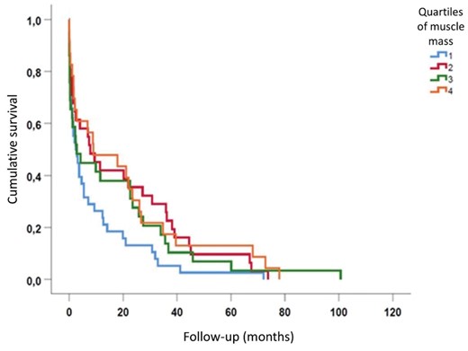
Cumulative survival of ESKD patients on hemodialysis divided by quartiles of muscle mass.


Comments