


Abstract
The retention of a large number of solutes that are normally excreted or metabolized by the kidney is responsible for the classical symptoms of haemodialysis (HD) patients. These molecules are defined as uremic toxins and can be classified into three groups: small water-soluble molecules, middle molecules and protein-bound toxins. Recently, efforts were put in order to develop dialysis membranes that allow the removal of large-middle molecules without clinically relevant albumin loss. These dialysers are the medium cut-off (MCO) membranes, which allow the removal of middle molecules up to approximately 50,000 Da. We performed a prospective, open-label, controlled, cross-over pilot study comparing expanded HD (HDx) with MCO membrane Theranova and conventional HD in 20 patients.
Ten patients underwent conventional HD high-flux dialyser and 10 patients underwent HDx for 3 months, later the patients switched and received the other treatment for a further 3 months. We then analyzed the pro-calcifying effect of patient uremic serum in a model of high-phosphate (Pi) induced calcification in vascular smooth muscle cells (VSMCs).
In this study each patient was the control of himself and, interestingly, we found a tendency of less pro-calcifying potential of serum from patients when treated with HDx, in comparison with HD (6.6±1.1, 5.6±0.6 and 6.7±1.0, OD/mg protein, baseline HD, 0-3 HDx and 3-6 HD, months, respectively; and 4.7±0.7 vs 3.2±0.2, OD/mg protein, 0-3 HD vs 3-6 HDx, months, respectively) (Figure). Investigating the major mechanisms involved in high-Pi induced calcium deposition, we found that serum from patients when treated with HDx induced less necrosis in VSMCs, compared with HD (3.2±0.8 vs. 0.9±0.1, necrosis enrichment factor, 0-3 HD vs. 3-6 HDx, months, respectively). Nevertheless, no differences were found between the different dialysis treatment in the serum potential to induce apoptosis and to modulate the expression of a panel of genes involved in VSMC osteoblastic-differentiation such as BMP2, RUNX2, osteocalcin, MGP, OPN, and elastin. In the effort to characterize the difference in uremic toxin profile during the two different dialysis treatments, we measured a panel of protein-bound uremic toxins which have been shown to promote vascular calcification and /or bone loss and shown a significant increased removal by HDx of 3-indoxyl sulfate (30.6±13.2 vs -17.1±10.2 Δ%; 0-3 HD vs 3-6 HDx, months; p<0.05; and -21.1±11.4 vs 54.4±33.2 Δ%; 0-3 HDx vs 3-6 HD, months; p<0.05), indole 3-acetic acid (43.1±16.9 vs -2.3±10.9 Δ%; 0-3 HD vs 3-6 HDx, months; p<0.05), CMPF (61.8±27.6 vs -16.1±9.6 Δ%; 0-3 HD vs 3-6 HDx, months; p<0.05), and kynurenine (44.2±12.9 vs -0.2±6.8 Δ%; 0-3 HD vs 3-6 HDx, months; p<0.05).
These preliminary data suggest a role for HDx in reducing the calcifying potential of uremic serum. Our findings are promising and they need to be confirmed in larger studies.
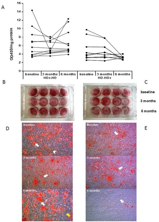
Calcification in VSMCs coultered with 10% uremic serum and challenged with 3.3 mM Pi for 3 days. Panel A left, B and D 0-3 months HDx and 3-6 months HD. Panel A right, C and E 0-3 months HD and 3-6 months HDx.
A Quantification of calcium deposition by Alizarin Red destaining. B-C Representative pictures of Alizarin Red staining in wells. D-E Ca deposition was visualized at light microscopic level (Alizarin Red staining). Magnification x200.


Comments