


Abstract
Treatment with high-volume HDF is associated with a better survival than standard HD. The underlying mechanism is unknown, but may depend on a lower level of intra-dialytic tissue damage. We compared tissue injury, as measured by the release of EVs during four dialysis treatment modalities.
Chronic hemodialysis (CHD) patients were included in an open, cross-over, randomized fashion (n=14). EV release was measured by a calibrated flow cytometer (Apogee A-60) after antibody labeling. EVs are defined as cell-derived particles with a diameter <1.000nm and a refraction index (RI) <1.42. Two HD modalities were studied: sHD, t dialysate 36,5°C (sHD) and cHD, t dialysate 35,5°C and two HDF modalities (t dialysate 36,5°C): lvHDF with a convection volume (CV) <15 L/session and hvHDF with a CV >23 L/session.To establish the cellular origin of EVs, EVs were labelled with (anti-) CD45 (leukocytes), CD62e (activated endothelial cells), CD62p (activated platelets), CD144 (vascular endothelial cadherin), CD61 (platelets), CD235a (erythrocytes) and connexin-43 (myocardial and endothelial cells), and with the general EV marker lactadherin. Differences between the 4 dialysis modalities were investigated by comparing pre- and post-dialysis and the delta (post- minus pre-dialysis) EV concentrations by Friedman’s test.
As shown in the figures below, CD61, lactadherin and CD144 increased during the 4 modalities (1=sHD,2=cHD,3=lvHDF,4=hvHDF). The overall increase (sum of all modalities) in connexin-43. In Table 1, the concentrations of EVs before- and after dialysis are shown with a p-value for the difference (post- minus pre-dialysis). No significant differences were found in pre- and post-dialysis and delta EV concentrations between the 4 dialysis modalities (data not shown).
From this analysis the following conclusions can be drawn: 1) except for CD62e, which declined, all measured EVs increased significantly during dialysis (Table 1). 2) Although not all EVs increased similarly during the 4 modalities (Fig 1), in terms of tissue damage no modality appears more advantageous than the other.
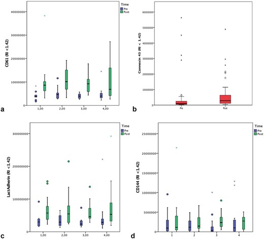
Absolute EV concentrations (mL-1) before and after dialysis: a:CD61, b:connexin-43, c:lactadherin, d:CD144. RI<1.42 (refraction index). Modality of dialysis (1= sHD, 2= cHD, 3= lvHDF, 4= hvHDF).
intradialytic EV changes: means of 4 modalities, delta’s and p-values post vs pre-dialysis
| EV concentration ( 107/mL) | Pre-dialysis | Post-dialysis | delta | p-value (post- vs pre) |
|---|---|---|---|---|
CD45 | 0.277 | 0.337 | 0.060 | <0.0005 |
| CD61 | 0.397 | 0.932 | 0.535 | <0.0005 |
| CD62p | 0.031 | 0.074 | 0.043 | <0.0005 |
| CD235a | 0.839 | 1.570 | 0.731 | <0.0005 |
| CD62e | 0.991 | 0.048 | -0.943 | 0.03 |
| CD144 | 0.021 | 0.163 | 0.142 | 0.03 |
| Connexin-43+ | 0.098 | 0.282 | 0.184 | 0.002 |
| Lactadherin | 2.826 | 5.176 | 2.35 | <0.0005 |


Comments