


Volume 71, Issue 2
April 2022
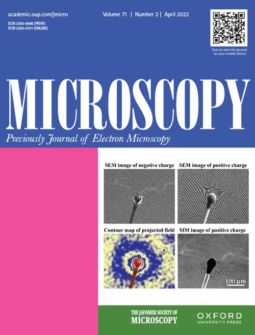
Cover image
Cover image
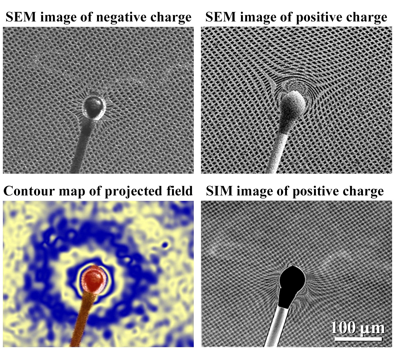
Lorentz Scanning Electron/Ion Microscopy: Observation and measurement methods for spatial electromagnetic fields were developed by using scanning electron/ion microscopes, in which a cross-grating was installed below the specimen. The specimens were observed under the infocus condition and the grating was simultaneously observed under the defocus condition. Projected electromagnetic fields around the specimen were reconstructed from the distorted cross-grating images in the similar way done by electron holography. The developed methods can be used in low and middle magnification and resolution ranges and are applicable to study their dynamics. Furthermore, these methods can be realizable in any electron/ion beam instruments because they are based on deflections due to Lorentz force for charged particle beams. Microscopy 71: 93–97.
EISSN 2050-5701
Issue navigation
Volume 71, Issue 2, April 2022
In This Issue
In This Issue
Microscopy, Volume 71, Issue 2, April 2022, Pages 1–3, https://doi.org/10.1093/jmicro/dfac013
Articles
Quadruple node of triple junctions of grain boundaries in a Eu2+-doped solid solution of the ions K+, Rb+, Cl− and Br−: an epifluorescence microscopy study using the doping ion as a fluorochrome
Microscopy, Volume 71, Issue 2, April 2022, Pages 77–86, https://doi.org/10.1093/jmicro/dfab047
Visualization of three-dimensional stigmoid body in FFPE and ultrathin sections of mouse
Microscopy, Volume 71, Issue 2, April 2022, Pages 87–92, https://doi.org/10.1093/jmicro/dfab052
Lorentz scanning electron/ion microscopy
Microscopy, Volume 71, Issue 2, April 2022, Pages 93–97, https://doi.org/10.1093/jmicro/dfab054
Study of high–low KPFM on a pn-patterned Si surface
Microscopy, Volume 71, Issue 2, April 2022, Pages 98–103, https://doi.org/10.1093/jmicro/dfab055
Correlative light and electron microscopy of poly(ʟ-lactic acid) spherulites for fast morphological measurements using a convolutional neural network
Microscopy, Volume 71, Issue 2, April 2022, Pages 104–110, https://doi.org/10.1093/jmicro/dfab058
Development of tilt-scan system for differential phase contrast scanning transmission electron microscopy
Microscopy, Volume 71, Issue 2, April 2022, Pages 111–116, https://doi.org/10.1093/jmicro/dfac002
Toward complex observation in electron microscopy using two-dimensional electron detector coupled with phase plate STEM
Microscopy, Volume 71, Issue 2, April 2022, Pages 117–123, https://doi.org/10.1093/jmicro/dfac004
Serial ultrathin sections to identify ultrastructural localization of GLUT1 molecules in vesicles in brain endothelial cells—correlative light and electron microscopy in depth
Microscopy, Volume 71, Issue 2, April 2022, Pages 124–131, https://doi.org/10.1093/jmicro/dfac005

