


Diatom test is known as a method that forensic scientists often used for examining diatoms, under the optical microscope, contained in major organs to determine whether the cause of death of immersion remains is drowning. The aim of this study was to examine whether SEM is useful for observing diatoms in cadaveric liver, which appears to be drowned. The pig liver was immersed with seawater, left at room temperature for three days. Ten gram of the liver was digested by three different methods; fuming nitric acid and sulfuric acid, fuming nitric acid and hydrogen peroxide, and proteinase K. After the digestive step, sample was diluted with pure water and passed through a cellulose filter. The filter was observed and compared under the optical microscope and SEM. As a result, SEM enabled detailed morphological observation of diatoms, some of which were difficult to observe by the optical microscope. In SEM observation, it is possible to discern even broken diatoms attached to organic materials, although manual SEM operation may take longer. As a future prospect, by developing auto-scan and image recognition by deep learning, it can be expected to increase its viability.
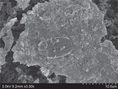
Diatom buried in residue, which was considered difficult to observe with an optical microscope.
