


Cover image

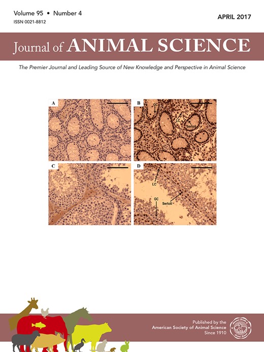
COVER ART: Immunolocalization of GATA-binding protein 4 (GATA-4) in the nuclei of Sertoli cells in a prepubertal bull (panels A and B) and a postpubertal bull (18 mo old; panels C and D). All sections shown are counterstained with hematoxylin and eosin. Panels A and C depict testicular sections in which GATA-4 blocking peptide was used to confirm the specificity of the primary antibody, and these sections appear identical to sections stained only with hematoxylin and eosin. Positive staining for GATA-4 is depicted in panels B and D by reddish-brown staining in Sertoli cell nuclei (Sertoli) but not germ cell nuclei (GC) within the seminiferous tubules. Leydig cell nuclei (LC) also stain positively for GATA-4 (panels B and D). For the enumeration of Sertoli cells (Table 2) in Exp. 2, only positively stained Sertoli cells in the cross section monolayer of a seminiferous tubules were quantified. Magnification bar on each micrograph represents 100 μm. For more information, see J. Anim. Sci. 95:1669–1679. doi:10.2527/jas.2016.1067

