-
PDF
- Split View
-
Views
-
Cite
Cite
Konstantinos P. Letsas, Gerasimos Gavrielatos, Michalis Efremidis, Stavros P. Kounas, Gerasimos S. Filippatos, Antonios Sideris, Fotios Kardaras, Prevalence of Brugada sign in a Greek tertiary hospital population, EP Europace, Volume 9, Issue 11, November 2007, Pages 1077–1080, https://doi.org/10.1093/europace/eum221
Close - Share Icon Share
Abstract
The purpose of the present study was to determine for the first time the prevalence of Brugada-type electrocardiographic (ECG) pattern (Brugada sign) in unselected individuals served by an urban Greek tertiary hospital during a 4-year time period.
Among 11 488 individuals (6640 males, 4848 females), 25 (23 males, 2 females, aged 36.8 ± 19.2 years) were found to display the Brugada sign (0.22%). Two cases exhibited the diagnostic type 1 ECG pattern (0.02%) and 23 subjects fulfilled the ECG criteria for type 2 or 3 patterns (0.2%). The incidence of Brugada sign was higher among men (0.34%) than in women (0.04%). Structural heart disease was established in four cases (one of them exhibiting a type 1 ECG pattern). Twenty-one individuals (19 males, 2 females, aged 29.7 ± 10.7 years) without structural heart disease displaying Brugada-type ECG features (4 cases with spontaneous or procainamide-induced type 1 ECG pattern) were subsequently selected and closely followed up for 24 ± 12 months. No mortality or life-threatening ventricular arrhythmias were recorded during this period.
The Brugada-type ECG pattern is infrequently seen in a Greek hospital-based population. All subjects with Brugada sign and structurally normal hearts displayed a benign clinical course without arrhythmic events during a relatively long follow-up period.
Introduction
The Brugada syndrome (BS) is an inherited channelopathy associated with a high propensity of polymorphic ventricular tachycardia, ventricular fibrillation, and sudden cardiac death in individuals with structurally normal hearts.1,2 The characteristic electrocardiographic (ECG) pattern referred as Brugada sign consists of complete or incomplete right bundle branch block and ST-segment elevation in leads V1 through V3.1,2 Mutations of the SCN5A gene encoding the α subunit of cardiac sodium channel have been reported as the genetic basis of the disease.3 The ECG features of BS could be intermittent, requiring a pharmacological challenge with a class I antiarrhythmic agent (ajmaline, flecainide, procainamide) to unmask the characteristic ST-segment elevation in right precordial leads.1,2
The frequency of Brugada sign varies significantly among ethnic populations, higher in south-eastern Asian countries, an event possibly ref lecting the geographical genetic distribution of the disease.1,2 A higher rate of arrhythmic events has been additionally demonstrated in Asian compared with European patients.4 The prognostic significance of Brugada sign remains a matter of considerable debate.1,5,6 No data regarding the incidence of Brugada-type ECG pattern exist in the Greek population. The aim of the present study was to determine the prevalence of Brugada sign in unselected individuals served by a Greek tertiary hospital. The prognostic significance of Brugada-type ECG pattern was additionally investigated.
Methods
Definition of Brugada-type ECG
Electrocardiograps were recorded at standard gain (10 mm/mV) and paper speed (25 mm/s). The diagnosis of Brugada sign was strictly based on the recommendations of the Second Consensus Conference on BS.1 According to this report, three types of ECG repolarization patterns in right precordial leads (V1–V3) have been recognized. Type 1 is diagnostic of BS and is characterized by a coved ST-segment elevation ≥2 mm followed by a negative T wave in more than one right precordial leads (Figure 1A). Type 2 ST-segment elevation displays a saddleback configuration with a high takeoff ST-segment elevation of ≥2 mm, a trough displaying ≥1 mm ST elevation, and either a positive or biphasic T wave (Figure 1B). Type 3 has either a saddleback or coved appearance with an ST-segment elevation of ≤1 mm (Figure 1C).

Examples of baseline ECG recordings with type 1 (A), type 2 (B), and type 3 (B) patterns of BS (leads V1–V3).
All ECGs were carefully evaluated by two independent investigators and the diagnosis of Brugada sign was made only when both investigators agreed on the classification of the ECG abnormalities.
Study population
The study population consisted of 11 488 unselected individuals (6640 males, 4848 females, aged 15–98 years) admitted to our Institution for different reasons (emergency department, out-patient clinics, preathletic screening, etc.) during a 4-year period. Our Institution is an urban tertiary referral health centre, the largest hospital in Greece, catering to an area of over 5 000 000 people. Subjects with ECG features of BS referred to our Institution for further evaluation were excluded from the study.
Individuals exhibiting the Brugada sign were carefully assessed regarding their medical history such as syncope or near syncope, chest discomfort, dyspnoea, palpitation, and their family history of sudden cardiac death, syncope, and heart diseases by means of questionnaires. Transthoracic echocardiography and ambulatory 24-h Holter recordings (Galix Biomedical Instrumentation, Miami, FL, USA) were performed to all subjects carrying the Brugada sign. The average heart rate, the standard deviation of all normal RR intervals (SDNN), and the standard deviation of the average normal RR intervals measured in successive 5-min segments (SDANN) were analysed. Baseline ECG parameters including QRS duration, PR interval, and corrected QT (QTc) interval (lead II) were also measured.
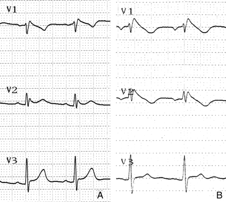
Subjects without structural heart disease exhibiting a type 2 or 3 ECG pattern underwent a sodium-channel-blocking test with procainamide (10 mg/kg). The diagnosis of BS was considered positive when the baseline type 2 or 3 ST-segment elevation converted to the diagnostic type 1 pattern (coved ST-segment elevation ≥2 mm) (Figure 2).1 Individuals without structural heart disease displaying the Brugada sign were subsequently selected, and closely followed-up for 24 ± 12 months.

Positive sodium-channel-blocking test with procainamide. The baseline type 2 ECG pattern with saddleback ST-segment configuration (A) was converted to the diagnostic type 1 pattern with coved ST-segment elevation (B).
The SPSS software package (Version 11.0 for Windows) was used for statistical analysis. Data are presented as mean ± SD. Comparisons between study groups were carried out using the Fisher's exact test. Differences with a P-value of <0.05 were considered as statistically significant.
Results
Among 11 488 unselected individuals, 25 (23 males, 2 females, aged 36.8 ± 19.2 years) were found to display the Brugada sign (0.22%). Two male subjects displayed the diagnostic type 1 ECG pattern (0.02%) and 23 subjects fulfilled the ECG criteria for type 2 or 3 patterns (0.2%). Of them, 7 were presented to the emergency department complaining palpitations or chest discomfort or dyspnoea, 15 were admitted for preathletic screening, and 3 were referred for cardiology consultation from the internal medicine and surgery departments. The incidence of Brugada sign was higher among men (0.34%) than in women (0.04%). The age-related prevalence of Brugada sign was estimated at 44% in subjects <30 years old, 40% in subjects 30–60 years old, and 16% in subjects >60 years old.
Structural heart disease was established in four individuals (one of them carrying a type 1 ECG pattern), and excluded from further evaluation. Three of them exhibited coronary artery disease and the other aortic valve stenosis. Echocardiography ruled out structural heart disease in the rest of the cases. Sodium-channel-blocking test with procainamide was performed in 18 out of 20 individuals with structurally normal hearts (two subjects denied) and converted the baseline type 2 or 3 ECG patterns to the diagnostic type 1 pattern in 3 cases.
Twenty-one individuals (19 males, 2 females, aged 29.7 ± 10.7 years) without structural heart disease displaying Brugada-type ECG features (4 cases with spontaneous or drug-induced type 1 ECG pattern) were subsequently selected and closely followed up for 24 ± 12 months. One of them was symptomatic (history of syncope) and received an implantable cardioverter defibrillator, whereas the others were asymptomatic.
Mean average heart rate (65.6 vs. 67.4 bpm), SDNN (102 ± 15 vs. 106 ± 29 ms), and SDANN (95 ± 20 vs. 100 ± 22 ms) values were found to share similar profiles in subjects with spontaneous or drug-induced type 1 ECG pattern and in subjects with type 2 or 3 ECG features, respectively (P > 0.05). Additionally, baseline ECG parameters including QRS duration (91.3 ± 11.5 vs. 90.8 ± 13.7 ms), mean PR (176.8 ± 26.5 vs. 173.1 ± 20.4 ms), and QTc (407.2 ± 42.2 vs. 414.3 ± 38.1 ms) intervals were found similar in both groups (P > 0.05). One case with spontaneous type 1 ECG pattern displayed a prolonged QTc interval (480 ms), whereas three other cases with type 2 ECG pattern exhibited a borderline QTc interval (range 433–440 ms).
No mortality or life-threatening ventricular arrhythmias were recorded during follow-up in subjects with Brugada-type ECG pattern.
Discussion
The ECG features of BS are dynamic and often concealed, and thus it is relatively difficult to estimate the true incidence of the disease in the general population.1 Furthermore, the occurrence of Brugada-type ECG pattern is strongly related to age. Previous studies have demonstrated that Brugada sign begins to appear during junior high school and increases until late adulthood.7,8
The frequency of Brugada sign varies significantly among ethnic populations. In Europe, among 35 309 individuals screened at a preventive medicine centre in France, 0.3% exhibited a spontaneous or drug-induced type 1 ECG pattern.9 In a different retrospective study from France including 1000 middle-aged subjects, the prevalence of Brugada sign was 6.1% (0.1% with type 1 ECG pattern).10 In a Finnish study including 2479 healthy male applicants and 542 healthy middle-aged subjects, 0.61% of the first population and 0.55% of the second population fulfilled the ECG criteria for type 2 or 3 of BS. Type 1 pattern was not seen in any subject (0%).11 The incidence of the Brugada-type ECG pattern in the general population of Southern Turkey was estimated at 0.48% (0.08% with type 1 ECG pattern).12 Viskin et al. showed that the prevalence of Brugada sign among healthy people in Israel is <0.5% (0% with type 1 ECG pattern).13
The incidence of Brugada sign has been found comparatively higher in south-eastern Asia and Japan. In a recent study including 3895 individuals from southern Iran, 2.56% displayed the Brugada sign. Of these, 0.54% exhibited a spontaneous or procainamide-induced type 1 ECG pattern.14 A community-based population study of 13 929 Japanese subjects demonstrated a 0.7% prevalence of Brugada-type ECG pattern (0.12% with type 1 ECG pattern). In male subjects, the Brugada sign was found in 2.14% (0.38% with type 1 ECG pattern).15 In a different study from the same country including 4788 subjects, the incidence of Brugada sign was 0.66%.16 On the contrary, Furuhashi et al. investigated 8612 Japanese subjects who underwent a health check up and found a very low incidence of Brugada-type ECG pattern (0.14%).17 In a hospital population study including 20 565 patients from Taiwan, the prevalence of Brugada sign was also found very low (0.13%).18
In USA, the frequency of Brugada-type ECG pattern was estimated at 0.43% (0.02% with type 1 ECG pattern) in a hospital population study including 12 000 non-cardiac patients.19 However, Patel et al. reported a very low incidence of Brugada sign (0.012%) among 162 590 unselected individuals served by a tertiary medical centre in USA.20
The present hospital population study determined the incidence of Brugada sign in Greek individuals at 0.22% (0.02% with spontaneous type 1 ECG pattern) which is lower compared with other European and Mediterranean countries (0.48–0.61%) and some south-eastern Asian countries (0.66–2.56%). We also showed that the frequency of Brugada sign is eight to nine times higher among men than in women. A noteworthy finding that needs further investigation was the overlap between Brugada-type ECG pattern and QTc interval prolongation. In our study, one symptomatic subject with spontaneous type 1 ECG pattern displayed a marked QTc interval prolongation, whereas three other asymptomatic cases with type 2 ECG features exhibited a borderline QTc interval. QT interval prolongation in patients with BS has been described in previous studies.21,22 A QTc >460 ms in V2 has been associated with an increased risk of arrhythmic events.22
The prognostic significance of Brugada sign remains a matter of considerable discuss. A recent meta-analysis showed that individuals with spontaneous type 1 ECG features exhibit a three- to four-fold increased risk of events compared with those with a drug-induced ECG pattern.4 On the contrary, type 2 or 3 ECG features in asymptomatic subjects have been considered as a normal variant rather than a specific warning sign of sudden cardiac death.11 In the current study, we closely followed up 21 subjects with Brugada sign for 24 ± 12 months. No mortality or life-threatening ventricular arrhythmias were observed in either symptomatic or asymptomatic cases. Symptomatic individuals exhibiting a spontaneously type 1 ECG pattern have an ∼six-fold increased risk of cardiac events, and according to the current recommendations they should receive an ICD without further evaluation.1,6 In contrast, previous studies have clearly showed that asymptomatic subjects exhibit a very low incidence of cardiac events ranging from 0 to 1.5%, a fact which is in accordance with our findings.9,11,14,15,18,23–26 Only one study have reported an 8% occurrence of cardiac events in initially asymptomatic subjects with Brugada-type ECG pattern.5 Therefore, asymptomatic subjects with Brugada sign are at low risk of developing life-threatening events.
In conclusion, the prevalence of Brugada-type ECG pattern in a Greek hospital-based population was found comparatively low. All subjects with structurally normal hearts and Brugada-type ECG pattern displayed a benign clinical course without arrhythmic events during a relatively long follow-up period. The presence of Brugada sign, mainly type 2 and 3 patterns, in asymptomatic individuals is very likely to represent a normal ECG variant.
Study limitations
Considering the dynamic nature of the ECG features, the true incidence of Brugada sign might have been underestimated. Furthermore, several conditions may simulate the Brugada-type ECG pattern.1,2 Although type 2 and 3 ECG patterns have precisely defined, the differentiation between the saddleback-type ST-segment elevation and the early repolarization syndrome is always susceptible to subjective bias. Our study included heterogeneous hospital population and not a community-based sample, and therefore may not reflect the real prevalence of Brugada sign in the general population. Finally, screening test for SCN5A gene mutations was not performed. However, the presence of SCN5A gene mutation is not related to the risk of events in BS.4
Conflict of interest: none declared.


