-
PDF
- Split View
-
Views
-
Cite
Cite
T. Rauwolf, M. Guenther, N. Hass, A. Schnabel, M. Bock, M.U. Braun, R.H. Strasser, Ventricular oversensing in 518 patients with implanted cardiac defibrillators: incidence, complications, and solutions, EP Europace, Volume 9, Issue 11, November 2007, Pages 1041–1047, https://doi.org/10.1093/europace/eum195
Close - Share Icon Share
Abstract
The present study evaluates the incidence of various complications in implanted cardiac defibrillators (ICD) therapy due to ventricular oversensing (VO) and its complications. From June 1998 to May 2005, we retrospectively screened 518 patients (1085.6 patient years) for the occurrence of VO episodes (441 male, 77 female). The overall incidence was 7.3% (n = 38) with inappropriate shock deliveries accounting for 2.3% (n = 12). All VO episodes were caused by either T-wave oversensing (n = 10), myopotentials (n = 8), electrode failure (n = 5), interference with electromagnetic fields (n = 3), double-counting (n = 4), pacemaker interactions (n = 2), or others (n = 2). There were five life-threatening events due to inappropriate ICD reaction. In eight (22%) cases, ICD reprogramming was able to avoid further oversensing episodes (e.g. adaptation of sensitivity, T-wave suppression feature), 13 (35%) patients had to undergo invasive procedures (e.g. electrode replacing) to suppress VO, 16 (43%) were told to avoid the trigger situation, and one demanded to deactivate all ICD therapies because of inappropriate shock delivery. Our data demonstrate that VO is a rare complication, but might lead to life-threatening events. In most cases, VO episodes could be prevented by appropriate ICD reprogramming or avoidance of the initiating trigger.
Introduction
In recent years, various multicenter, prospective studies have shown a benefit in survival after implanted cardiac defibrillators (ICD) implantation in patients with impaired left ventricular function1–4 or other cardiac diseases, such as Brugadás syndrome, long QT syndrome, or arrhythmogenic right ventricular dysplasia (ARVD).5,6 However, accurate and effective ICD function depends on correct sensing of intracardiac electrical activity and their appropriate interpretation by individual decision algorithms.7 Oversensing is a well-known phenomenon potentially leading to ICD malfunctioning by interfering with intracardiac signals and usually is related to myopotentials, T-waves, electrical spikes of an additionally implanted pacemaker or other technical devices (i.e. microwaves, stimulation therapy in physiotherapy, and electric coagulation8–11). The ICD system often misinterprets these cardiac/non-cardiac potentials as a malignant arrhythmia and delivers inappropriate shock therapy, which in many patients is an extremely uncomfortable event and greatly affecting quality of life. In some cases, inappropriate therapy delivery is described as potentially life threatening,12 especially when the shock falls in the vulnerable phase of QRS complex, triggering ventricular tachycardia (VT) or ventricular fibrillation (VF).
The aim of the present study was to evaluate in a large cohort of 518 ICD patients the incidence and various types of ventricular oversensing (VO), the occurrence of inappropriate shock deliveries as well as the frequency of complications requiring invasive procedures to solve VO during a long-term follow-up.
Materials and methods
Patient population
All 518 consecutive patients, who received an ICD implantation in our clinic between June 1998 and May 2005, were included. The individual ICD devices (including all ICD exchanges, e.g. because of low battery status) were provided by Biotronik (Berlin, Germany; n = 113), ELA (Cedex, France; n = 2), St Jude Medical (St. Paul, USA; n = 2), Guidant/CPI (St. Paul, USA; n = 524), and Medtronic (Minneapolis, USA; n = 160). Regular follow-up visits were usually scheduled in 3 months intervals in our outpatient clinic. During each device interrogation, all important parameters of ICD were checked (e.g. battery status, lead and shock impedance, pacing thresholds, and atrial and ventricular sensing) as well as the ICD memory function screened for potential episodes of VO. Cardiological examination (echocardiography and stress tests) was performed every 12 months based on the clinical judgement of the physician. Oversensing provocation manoeuvres or X-ray examination were performed, if an electrode lead failure (changes in impedance and unclear stored EGM) was assumed. Altogether 1085.6 patient-years were documented and the VO problems were categorized to the origin of noise signal, including T-wave oversensing, myopotentials, electrode failure, interference with electromagnetic fields, double-counting of broad QRS complexes, pacemaker interactions, and others.
Statistical analysis
All data and statistics are reported as mean ± standard deviation (n± SD) for continuous normally distributed data or as median (25–75%) for not normally distributed variables, respectively. Categorical variables are shown as number (%). P-values below 0.05 were considered significant.
Patients' characteristics
Five hundred and eighteen consecutive patients (i.e. 1085.6 patient-years) with an ICD implantation were studied retrospectively in respect of the occurrence of VO episodes and the incidence of inappropriate therapy deliveries. The median of follow-up was 1.64 years per patient (25–75%: 0.52–3.39 years). As shown in Table 1, the mean left ventricular ejection fraction (LVEF) was 31.9 ± 11.2% at the time of implantation, 312 (60.2%) patients had ischaemic cardiomyopathy, 163 (31.5%) suffered from non-ischaemic (dilatative) cardiomyopathy, and 43 (8.3%) had other forms of heart disease (i.e. ARVD, long QT syndrome).
| . | All patients . | Male . | Female . | P-value . |
|---|---|---|---|---|
| Patients | 518 | 441 | 77 | |
| Patient-years | 1085.6 | 938.5 | 147.1 | |
| Age (years) | 61.2 ± 11.0 | 61.4 ± 10.3 | 60.8 ± 12.0 | n.s. |
| Abdominal ICD | 11 | 9 | 2 | n.d. |
| LVEF (%) at the time of implantation | 31.9 ± 11.2 | 31.8 ± 10.9 | 33.0 ± 13.3 | n.s. |
| ICM | 312 | 278 | 34 | n.s. |
| LVEF (%) | 32.5 ± 9.6 | 31.8 ± 10.9 | 33.0 ± 13.3 | |
| DCM | 163 | 129 | 34 | |
| LVEF (%) | 25.6 ± 7.9 | 25.6 ± 7.8 | 26.0 ± 8.2 | n.s. |
| ARVD | 3 | 2 | 1 | |
| LVEF (%) | 51.7 ± 2.9 | 52.5 ± 3.5 | 50 | n.d. |
| Long QT syndrome | 3 | 0 | 3 | |
| LVEF (%) | 61.7 ± 2.8 | 61.7 ± 2.8 | n.d. | |
| Other aetiology | 9 | 7 | 2 | |
| LVEF (%) | 44.4 ± 15.9 | 38.6 ± 12.9 | 65 ± 0 | n.d. |
n.s., not significant; n.d., not determined due to low number.
| . | All patients . | Male . | Female . | P-value . |
|---|---|---|---|---|
| Patients | 518 | 441 | 77 | |
| Patient-years | 1085.6 | 938.5 | 147.1 | |
| Age (years) | 61.2 ± 11.0 | 61.4 ± 10.3 | 60.8 ± 12.0 | n.s. |
| Abdominal ICD | 11 | 9 | 2 | n.d. |
| LVEF (%) at the time of implantation | 31.9 ± 11.2 | 31.8 ± 10.9 | 33.0 ± 13.3 | n.s. |
| ICM | 312 | 278 | 34 | n.s. |
| LVEF (%) | 32.5 ± 9.6 | 31.8 ± 10.9 | 33.0 ± 13.3 | |
| DCM | 163 | 129 | 34 | |
| LVEF (%) | 25.6 ± 7.9 | 25.6 ± 7.8 | 26.0 ± 8.2 | n.s. |
| ARVD | 3 | 2 | 1 | |
| LVEF (%) | 51.7 ± 2.9 | 52.5 ± 3.5 | 50 | n.d. |
| Long QT syndrome | 3 | 0 | 3 | |
| LVEF (%) | 61.7 ± 2.8 | 61.7 ± 2.8 | n.d. | |
| Other aetiology | 9 | 7 | 2 | |
| LVEF (%) | 44.4 ± 15.9 | 38.6 ± 12.9 | 65 ± 0 | n.d. |
n.s., not significant; n.d., not determined due to low number.
Episodes of VO were documented in 38 (7.3%) patients during follow-up, whereas 12 (2.3%) patients suffered from inappropriate anti-tachycardic ICD therapy, 37 times recorded as inappropriate shock deliveries and two times as inappropriate ATP (Table 2). There were five episodes of potentially life-threatening conditions due to VO, i.e. one patient received an inappropriate ICD shock while swimming through an electrically opening pool door. No documented ICD malfunctions due to VO resulted in fatal outcome. However, we have to confirm that no final read out of ICD memory was taken in 96 cases post-mortem.
| Patients with VO | 38 (7.3%) |
| Patients with inappropriate shocks | 12 (2.3%) |
| Number of life-threatening episodes due to VO | 5 |
| Sum of inappropriate shocks caused by VO | 37 |
| Documented fatal complication | 0 |
| Patients with VO | 38 (7.3%) |
| Patients with inappropriate shocks | 12 (2.3%) |
| Number of life-threatening episodes due to VO | 5 |
| Sum of inappropriate shocks caused by VO | 37 |
| Documented fatal complication | 0 |
| Patients with VO | 38 (7.3%) |
| Patients with inappropriate shocks | 12 (2.3%) |
| Number of life-threatening episodes due to VO | 5 |
| Sum of inappropriate shocks caused by VO | 37 |
| Documented fatal complication | 0 |
| Patients with VO | 38 (7.3%) |
| Patients with inappropriate shocks | 12 (2.3%) |
| Number of life-threatening episodes due to VO | 5 |
| Sum of inappropriate shocks caused by VO | 37 |
| Documented fatal complication | 0 |
The underlying mechanisms of VO episodes were characterized as demonstrated in Table 3.
Mechanism of ventricular oversensing and number of inappropriate ATP/shock deliveries
| Mechanism of oversensing . | Number of patients . | Accumulated inappropriate shock/ATP deliveries . |
|---|---|---|
| Myopotentials | 8 | 8 |
| Electrode failure | 5 | 6 |
| Electromagnetic field interference source inclusive medical interference source | 4 | 4 |
| T-wave oversensing | 10 | 3/1 |
| Pacemaker interactions | 2 | 5 |
| Double-count mechanism | 4 | 8/1 |
| Not classified | 1 | 1 |
| Patients with more than one mechanism of oversensing | 4 | 2 |
Mechanism of ventricular oversensing and number of inappropriate ATP/shock deliveries
| Mechanism of oversensing . | Number of patients . | Accumulated inappropriate shock/ATP deliveries . |
|---|---|---|
| Myopotentials | 8 | 8 |
| Electrode failure | 5 | 6 |
| Electromagnetic field interference source inclusive medical interference source | 4 | 4 |
| T-wave oversensing | 10 | 3/1 |
| Pacemaker interactions | 2 | 5 |
| Double-count mechanism | 4 | 8/1 |
| Not classified | 1 | 1 |
| Patients with more than one mechanism of oversensing | 4 | 2 |
| Mechanism of oversensing . | Number of patients . | Accumulated inappropriate shock/ATP deliveries . |
|---|---|---|
| Myopotentials | 8 | 8 |
| Electrode failure | 5 | 6 |
| Electromagnetic field interference source inclusive medical interference source | 4 | 4 |
| T-wave oversensing | 10 | 3/1 |
| Pacemaker interactions | 2 | 5 |
| Double-count mechanism | 4 | 8/1 |
| Not classified | 1 | 1 |
| Patients with more than one mechanism of oversensing | 4 | 2 |
T-wave oversensing
The most frequent oversensing mechanism was observed as T-wave oversensing in 10 patients.13,14 This type of VO is caused by sensing T-waves as additionally QRS complexes. So ICD’s count ventricular frequency inadequately high and an inappropriate shock delivery is possible.15–17 Three inappropriate shocks and one ATP were delivered in observed periods. In three patients (30%) of the present study group, a T-wave suppression feature was effective to avoid oversensing. In three patients (30%), a replacement of pace/sense electrode was required. Figure 1 shows a typical example for T-wave oversensing.
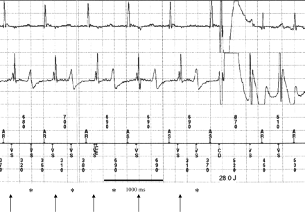
Ventricular oversensing due to T-wave oversensing, resulting in inappropriate shock therapy. Correctly sensed R-waves are marked with downward arrow, T-wave oversensing (misinterpreted as ventricular exitation) is marked with asterisk.
Oversensing caused by myopotentials
In the present study, eight (1.5%) patients were noticed with VO due to myopotentials.8,18 This type of VO in ICD therapy is a rare complication caused by the exclusively used bipolar leads.19–22 All registered episodes were related to contraction of the respiratory, trunk, or upper arm musculature. Whereas in three patients, the implantation of a new pace/sense lead was necessary, reprogramming of the ICD with changing the ventricular sensitivity value was sufficient to avoid further oversensing episodes. All other cases only showed short episodes without therapeutical consequence. Figure 2A shows IEGM in a case of myopotential oversensing.
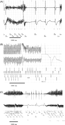
(A) Myopotential oversensing caused by trunk and upper arm musculature while rising up after echocardiography. Myopotential oversensing was reproducible during the following provokation manoeuvre. (B) Oversensing due to external electromagnetic field. Inadequate shock delivery caused by electromagnetic field (electromagnetic field transmitter)—noise signals induced in atrial and ventricular channel misinterpreted as ventricular fibrillation, detailed description see text. (C) Inappropriate shock delivery caused by oversensing due to alternating current stimulation therapy in a patient with myopathia.
Oversensing caused by lead failure/lead fracture and insulation defect
Five patients suffered from VO due to electrode failure and consecutive noise sensing with inadequate therapy delivery.23–25 In all patients, an electrode replacement was required. Lead failure or fracture can imitate all other types of oversensing. An essential role in preventing complications caused by electrode failure is to register impedance of the lead exactly. All cases of unexpected changes in lead impedance require precise physical examination, provocation manoeuvres, and chest X-ray examination as standard diagnostic procedures.
Oversensing caused by double-counting
Double-counting was recorded in four patients, leading to eight inadequate shocks in two patients and short episodes without therapy delivery in the others. In our study group, we found two underlying mechanisms of this VO type. In one patient with an older biventricular system, the ventricular channel of the left and right ventricular leads was connected by a Y-adapter to the device, the ventricular potential was sensed twice because of the underlying intraventricular conduction delay. In the other three patients, double-counting was due to a broad QRS complex. In these cases, the ICD detects one QRS complex twice and misinterprets a normal sinus rhythm or a slow VT as malignant tachyarrhythmia. Figure 3 shows a double-count mechanism in a patient with a running, haemodynamically tolerated slow VT (CL 590 ms). In this patient, a replacement of pace/sense lead was performed.
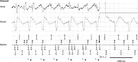
Double-counting while incessant (haemodynamically tolerated) slow VT (CL 590 ms), every QRS complex is counted twice as marked with asterisk and x. The delivery of an inappropriate shock delivery is marked with downward arrow.
Oversensing caused by alternating current application and electromagnetic fields
Three patients experienced VO according to electromagnetic field interference with the ICD device by inducing an alternating current in the sensing electrode or to alternating current, respectively. In one patient, microwave energy was applied for pain relief at the skin close to the ICD pocket. The signals from electromagnetic field transmitter were detected from the atrial and ventricular channel of the ICD, misinterpreted as VF inducing an inappropriate shock delivery (Figure 2B). The second patient had VO while swimming through an electrically opening pool door. In another case, the inadequate shock was due to an alternating current application during physiotherapy session using electric stimulation of the skin (Figure 2C).
Interactions between pacemaker and implanted cardiac defibrillators
Six patients had an ICD and an additionally implanted pacemaker. There are different reasons for these double device implantations:
first generation of ICD’s with large volume were implanted abdominally;
first generation ICD’s were only available as single-chamber devices;
pre-existing biventricular pacemaker for CHF therapy;
In the present study, two of six (33.3%) patients were noticed with episodes of such interaction. One patient suffered from a life-threatening condition due to an interaction of the ICD and the pacemaker, resulting in haemodynamically not tolerated bradycardia for some seconds resulting in a circulatory collapse and unconsciousness (Figure 4). Initially, the pacemaker (ELA, Talent DR, DDD 55–130) showed a typical ventricular undersensing while an incessant slow VT (CL 480 ms). Caused by inappropriate ventricular stimulation, slow VT was accelerated to polymorphic VT/VF. This resulted in an adequate and effective shock delivery. However, after shock delivery, the pacemaker was ineffective in stimulation (exit block) and simultaneously the ICD interpreted pacemaker spikes as normal QRS complexes—so the pacemaker function the ICD (VVI 30) was inadequately inhibited. A haemodynamically not tolerated bradycardia with slow ventricular rhythm was the consequence.
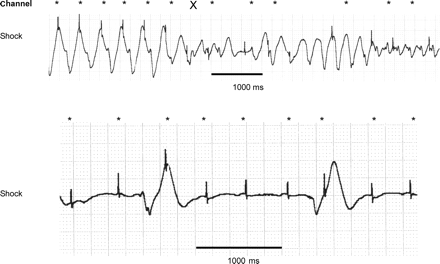
(A) Intermittent pacemaker undersensing while running slow VT (pacemaker spikes marked with asterisk), acceleration to VF due to pacemaker (marked with X). (B) Exit block of pacemaker after shock delivery, pacemaker (DDD 55–130) spikes are misinterpreted as QRS complexes by implanted cardiac defibrillators, resulting in haemodynamically not tolerated bradycardia (slow ventricular rhythm).
Another patient received inappropriate shock delivery while interrogation (Figure 5). A magnet test activated the ‘DOO 85’-Modus of the additionally implanted pacemaker system (Medtronic Thera DR, DDD 30–110) with unipolar atrial lead and bipolar ventricular lead.
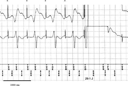
Interaction of dual chamber pacemaker and implanted cardiac defibrillators (ICD) DOO 85-Modus of implanted pacemaker (Medtronic TheraDR) while magnet test, atrial unipolar spikes are marked with asterisk, bipolar ventricular spikes are not sensed by ICD, inappropriate shock delivery triggered by misinterpreting atrial spikes.
Discussion
The present study was performed to analyse in a large cohort of 518 ICD patients the incidence and different mechanisms of VO during a long-term follow-up (1085.6 patient-years), including complications such as inappropriate shock deliveries as well as necessary invasive procedures to solve the underlying problem.
According to various multicenter, prospective studies showing a benefit in survival for patients with severe impaired left ventricular function, ICD implantation currently is recommended in different cardiac diseases as an effective, efficient, and save treatment option.4,5,28 Nevertheless, ICD therapy might be associated with technical/signalling faults beyond the well-known peri-interventional complications at the implantation site such as haematoma, bleeding, or infection.29 The 7.3% incidence of VO in our patient cohort is comparable with previously published data in smaller and selected patient groups.11,30 Only few patients (2.3%) needed an electrode replacement to solve the oversensing problem. In most of the cases, VO episodes could be avoided by reprogramming the ICD (e.g. T-wave suppression function), changing the ventricular sensitivity value, or by the renewed instruction the patient to omit potential ICD interference sources, i.e. electromagnetic fields. Despite the risk for potentially life-threatening conditions due to VO, no documented ICD malfunctioning resulted in fatal outcome.
However, 96 patients died in study interval had no read out of ICD memory post-mortem. Therefore, no reliable information about fatal complications potentially depending on inappropriate ICD therapy is available. Fatal complications due to inappropriate ICD therapy are described in some case reports13,15,31 but usually are seldom.
Lead failure is another mechanism described in the literature, which may cause fatal complications.23,32 A life-threatening situation caused by lead failure was not noticed. Ellenbogen et al.25 recently reported that special attention should be applied to older lead types. Unexpected changes in impedance, threshold, or unexpected noise sensing may precede complications. Advanced monitoring with provocation manoeuvres and chest X-ray are standard control procedures in all suspect cases.
Special attention is required in ICD patients with additionally implanted pacemaker systems. These devices can interfere significantly. Today, the combination of ICD and additionally implanted pacemaker is rare because all current ICD devices can be implanted subclavicular and obtain all pacemaker functions. Only a few patients with older abdominal implanted ICD devices or additionally implanted pacemakers are affected by device interactions. We confirm the results of the study group of Calkins et al.33 that interference of ICD and pacemaker is common and could be avoided in many cases by careful programming. However, in current guidelines for ICD and pacemaker implantation and follow-up, there is no information how to handle this special patient group in this double device situation.34 In such situations, the authors recommend to a careful assessment for an explantation of the pacemaker, when a sufficient ICD device is present.
Study limitations
The present study was retrospective in design and monocentric. However, all data and ICD memory episodes were evaluated by two independent cardiologists experienced in pacemaker and ICD interrogation.
Conclusions
In summary, VO is a rare cause of ICD malfunction and cannot be attributed as a specific problem to any of the manufacturer of ICD devices used. In most cases, oversensing does not result in severe complications and usually is resolved by non-invasive manoeuvres. Special attention has to be applied to patients with older types of leads and/or additionally implanted pacemaker. A sufficient instruction about behavioural patterns should be given to all ICD patients, to avoid oversensing episodes.
Conflict of interest: none declared.
Funding
No financial support was received for the present study.
Reference
Author notes
† Both authors contributed equally.


