


Abstract
We report a 48-year-old woman with manifest Wolff–Parkinson–White (WPW) syndrome and a long QT interval in her electrocardiogram, who represented the marked prolongation of QT interval after the successful catheter ablation for the accessory pathway. The increase in QT interval was completely restored to the baseline level 2 days after the procedure. Thus, the change in QT interval should be evaluated with particular caution after catheter ablation for WPW syndrome patients.
A 48-year-old female with manifest Wolff–Parkinson–White (WPW) syndrome without any family history of long QT syndrome was referred for the curative treatment to our institution. Before the ablation therapy, the patient exhibited long QT (Figure 1A, QTc interval; 554 ms). After the successful catheter ablation for left lateral accessory pathway, the electrocardiography demonstrated the marked prolongation of QT interval (QTc interval; 688 ms) even without any antiarrhythmic drug (Figure 1B). She had no symptom or malignant ventricular arrhythmias after the procedure. Laboratory data showed normal findings. The increase in the QT interval was restored to the baseline level 2 days after the catheter ablation (Figure 1C). Previous studies have shown that the QT interval was prolonged by a long period of pacing or ventricular pre-excitation,1,2 possibly in part by alterations of cardiac potassium channel functions. We carefully checked the QT interval after catheter ablation in this case, and found a transient marked prolongation of the QT interval after the procedure. We speculated the QT prolongation observed in this patient was due to the similar mechanism to that of cardiac memory or the above-mentioned previous reports showing repolarization abnormalities after pacing or ventricular pre-excitation.1,2 Thus, the change in QT interval should be evaluated with particular caution after catheter ablation for WPW syndrome patients.
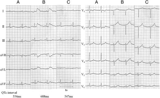
Twelve-lead electrocardiography before (A), immediately after catheter ablation for Wolff–Parkinson–White syndrome (B), and 2 days after the procedure (C). The increase in the QT interval observed immediately after the catheter ablation was restored to the baseline level 2 days after the procedure. The values of QTc interval in lead V2 were shown.
Conflict of interest: none declared.
