-
PDF
- Split View
-
Views
-
Cite
Cite
Eric Chong, Shih-Lin Chang, Shih-Ann Chen, Pulmonary vein isolation in a patient with dextrocardia, EP Europace, Volume 14, Issue 12, December 2012, Page 1725, https://doi.org/10.1093/europace/eus325
Close - Share Icon Share
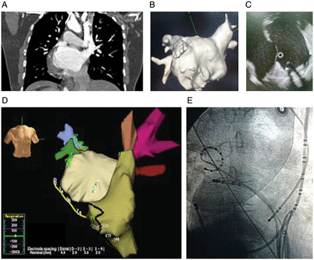
A 61-year-old lady with known dextrocardia and paroxysmal atrial fibrillation (AF) was referred for ablation. Computed tomography (CT) of the thorax (frontal plan as shown in Figure 1A) and CT geometry reconstruction (Figure 1B) were performed prior to ablation. Intracardiac echocardiography (Figure 1C) showed a secondum atrial septal defect (ASD). Atrial trans-septal assess was achieved through the ASD. Three-dimensional mapping of the atria (Figure 1D) using Ensite NavX system (St Jude Medical) showed complete reversal of the left and right atrium and pulmonary veins. Pulmonary vein isolation was performed successfully in the reverse manner (Figure 1E). The procedural time was 140 min and fluoroscopy time was 42.5 min. The patient tolerated the procedure well and there were no complications. This case demonstrated the complete reversal of cardiac anatomy during AF ablation.

Conflict of interest: none declared.


