


Cover image
Tako-tsubo cardiomyopathy, acute coronary syndrome, or both?
Stefano Svab, Elena Pasotti, Tiziano Moccetti, and Giovanni B. Pedrazzini*
Cardiocentro Ticino, Lugano, Switzerland
* Corresponding author. Tel: +41 918053178, Fax: +41 918053173, Email: [email protected]
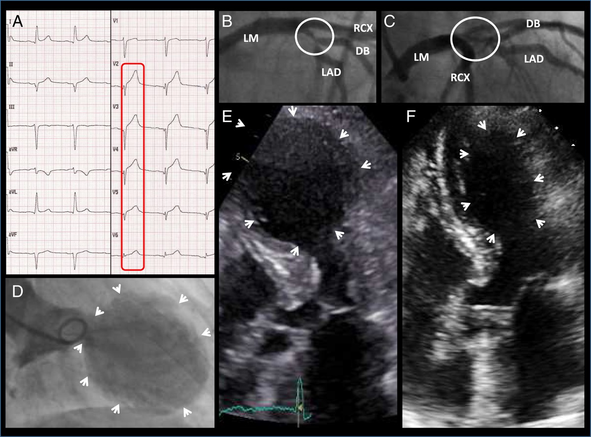
A 76-year-old Caucasian woman known for hypertension and former cigarette smoking was admitted for acute dyspnoea and chest pain with 12-lead ECG highly evocative for sub-acute anterior myocardial infarction (Panel A). Based on the urgent coronary angiogram showing a complex left anterior descending (LAD) artery proximal bifurcation lesion (Medina 1/1/1) with preserved peripheral flow (Panels B and C) and only a slight increase in troponin I (maximal value 5.5 μg/L, normal value <0.9 μg/L), the decision was taken to submit the patient to accelerated surgical revascularization. Unexpectedly, both the ventriculogram (Panel D) and the initial transthoracic echocardiography (Panel E) showed a severe antero-apico-basal akinesia, whereby surgical intervention was postponed. Based on the complete normalization of the LV function 1 week after the acute event (Panel F), we hypothesized an apical ballooning syndrome in a patient with concomitant severe LAD lesion most probably corresponding to an acute coronary syndrome. In the current literature it is still a matter of debate whether both pathologies might coexist simultaneously. This case strongly supports the hypothesis of a possible cohabitation.
Panel A. ECG at admission, showing ST elevation in anterior leads. Panels B and C. LAO cranial and RAO cranial angiographic views, respectively, showing a complex LAD proximal bifurcation lesion with preserved flow (Supplementary material online, Video S2 and S3). Panel D. Ventriculogram showing severe antero-apico-basal akinesia (Supplementary material online, Video S1). Panel E. Transthoracic echocardiogram at admission confirming severe antero-apico-basal akinesia (Supplementary material online, Video S4). Panel F. Transthoracic echocardiogram performed 1 week later, showing a normalization of LV function (Supplementary material online, Video S5). LM, left main artery; LAD, left anterior descending artery; DB, diagonal branch; RCX, circumflex artery.
Supplementary material is available at European Heart Journal online.
Volume 39, Issue 2, 07 January 2018
Special Issue on the 2017 ESC Guidelines for the management of acute myocardial infarction in patients presenting with ST-segment elevation
Issue @ A Glance
ST-segment elevation myocardial infarction: the new ESC Guidelines
CardioPulse
The 2017 ESC STEMI Guidelines
‘Ten Commandments’ of the 2017 ESC STEMI Guidelines
Dr David G. Harrison
European Sudden Cardiac Arrest network: towards Prevention, Education and New Effective Treatments (ESCAPE-NET): A major European Horizon 2020 project focused on cardiac arrest
Ferrara III
Clinical Research
Acute coronary syndromes
Patients with acute myocardial infarction and non-obstructive coronary arteries: safety and prognostic relevance of invasive coronary provocative tests
Editorial
Provocative tests for coronary artery spasm in MINOCA: necessary and safe?
Interventional cardiology
Long-term survival and causes of death in patients with ST-elevation acute coronary syndrome without obstructive coronary artery disease
EHJ Brief Communication
Acute coronary syndromes
A single-chain antibody-CD39 fusion protein targeting activated platelets protects from cardiac ischaemia/reperfusion injury

Editorial
Targeted antiplatelet therapy: novel treatment options for ischaemic heart disease


