


Cover image
Detection of pulmonary arteriovenous fistula with three-dimensional computed tomographic angiography
Chengming Fan1†, Zijing Zhou2†, Ni Yin1, and Jinfu Yang1*
1Department of the Cardiovascular Surgery, the Second Xiangya Hospital, Central South University, Changsha, China; and 2Department of Respiratory Medicine, the Second Xiangya Hospital, Central South University, Middle Renmin Road 139, Changsha 410011, China
*Corresponding author. Tel: +86 731 85296107, Fax: +86 731 85296606, Email: [email protected]
†These authorscontributed equally to this work.
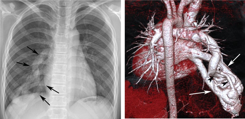
A 13-year-old boy presented with mild exercise-induced shortness of breath; he reported being otherwise healthy. Physical examination revealed a reduced arterial oxygen saturation level (82%) and mild cyanosis with clubbed fingers and toes. Chest radiography showed a tubular opacity in the right lower pulmonary lobe (Panels A and B, arrows). A computed tomography was performed and revealed a complex vascular structure in the right lower lobe (Panel C), and three-dimensional computed tomography angiography (Panel D, see Supplementary material online, Video) revealed an arteriovenous fistula with two feeding arteries (arrowheads) and two draining vein (arrows). Selective pulmonary angiography confirmed this diagnosis. The right lower pulmonary lobe was surgically removed, because the feeding arteries were not suitable for occlusion. Subsequently, the patient's arterial oxygen saturation level returned to normal (99%), and he is in good condition.
Supplementary material is available at European Heart Journal online.
Funding
Funding to pay the Open Access publication charges for this article was provided by the corresponding author.

