


Abstract
Patent foramen ovale (PFO) is one of the causes of cardioembolism and closure of PFO is recommended by the current guidelines in patients with recurrent stroke. Transoesophageal echocardiography (TEE) using bubble-contrast study is the gold standard imaging modality for the assessment of interatrial septum. Upper-extremity veins are the most common way of injection, however, the presence of Eustachian valve and flow dynamics when bubble-contrast injection performed via upper extremity veins limits the assessment of interatrial septum in several cases. In this study, we aimed to compare the efficacy of bubble-contrast study between upper extremity injection and lower extremity injection.
Patients with a suspicion of cardioembolism who were undergoing TEE study were included in this study. After routine assessment of cardiac structures, the bubble-contrast study was performed using agitated saline from both upper-extremity vein and lower-extremity vein with Valsalva manoeuvre. Right-to-left shunt and numbers of bubbles transmitted from the septum were recorded.
We prospectively included 45 patients and 21 PFOs were detected. There were 9 patients with prominent Eustachian valve and in 6 patients Eustachian valve hampered the complete opacification of the right atrium. In 3 patients flow from the superior vena cava was directed towards the tricuspid valve and hampered the complete opacification. Among 21 patients with PFO, in 6 patients right-to-left shunt was not observed when agitated-saline was injected via the upper-extremity vein, however, the shunt was observed when the agitated-saline was injected via the lower-extremity vein. In 14 patients amount of bubbles passing through the interatrial septum were significantly higher when the injection was performed via the lower-extremity vein especially in patients with prominent Eustachian valve.
Our preliminary results indicated that compared to upper-extremity veins, injection via the lower-extremity veins provides better opacification of right atrial septum and assessment of interatrial septum. Therefore, injection through the lower-extremity veins would be the preferred choice particularly in patients with prominent Eustachian valve or downward directed flow from the superior vena cava.
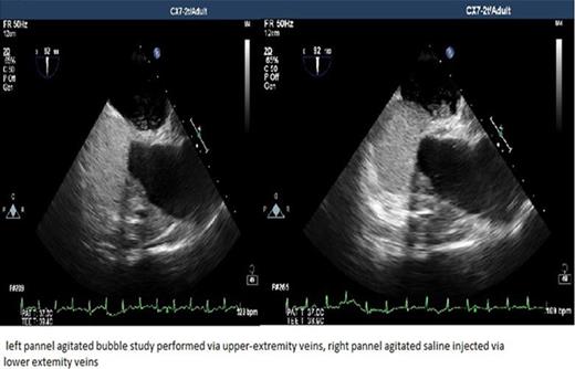
Figure 1
Type of funding source: None

