


Abstract
IgG4-related disease (IgG4-RD) is a relapsing-remitting immune-mediated condition that affects multiple organ systems. Case reports of suspected cardiovascular involvement in IgG4-RD have emerged though no study has systematically assessed the cardiovascular phenotype of IgG4-RD using cardiac magnetic resonance (CMR) imaging.
We systematically assessed patients with IgG4-RD using CMR to identify the cardiovascular manifestations, and compared them to controls.
We recruited 11 patients with histologically-confirmed IgG4-RD (6 female, 61±11 years, 9 with active disease (8 with pancreatic involvement, 3 parotid, 5 bile ducts, 5 kidneys and 3 cardiovascular)). Patients underwent CMR at 1.5T including cine, myocardial tagging, native T1-mapping, late gadolinium enhancement (LGE), extracellular volume (ECV) and quantitative stress perfusion mapping. Results were compared to 10 healthy controls with no cardiac disease (50% female, 35±8 years). Results are presented as mean ± standard deviation (SD) unless otherwise stated.
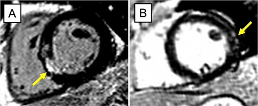
Patients with IgG4-RD had similar cardiac geometry to the reference group (Table 1), with similar biventricular and bi-atrial volumes. However, despite similar biventricular ejection fractions compared to controls (patient LVEF: 55-67%, RVEF: 51-65%), IgG4-RD patients showed significantly reduced global longitudinal strain (GLS) (-16.8 ± 2.3% vs -18.7 ± 1.6%, p=0.045). Furthermore, 64% (7/11) of IgG4-RD patients showed LGE, 6 of whom showed non-ischaemic patterns (Figure 1). Male IgG4-RD patients (n=5) as a group showed a significantly higher average myocardial T1 value relative to the reference group, with 3/5 male patients having an abnormally high myocardial T1 (above the 2SD limit of normal). Female IgG4-RD patients (n=6) had similar and normal myocardial T1 values to the reference group. ECV did not significantly differ between the two groups. IgG4-RD patients showed a significantly reduced global myocardial perfusion reserve (MPR), including two patients with a qualitative inducible perfusion defect. Only 3 out of the 11 IgG4-RD patients had a completely normal CMR.

Author notes
Funding Acknowledgements: Type of funding sources: Other. Main funding source(s): University of Oxford Medical Sciences Division Pump-Priming Grant

