


Abstract
Right ventricular (RV) dysfunction in sarcoidosis is associated with adverse outcomes. Assessment of RV function by conventional transthoracic echocardiography (TTE) is challenging due to the complex RV geometry. Knowledge-based reconstruction (KBR) combines TTE measurements with three-dimensional coordinates to determine RV volumes.
The aim of this study was to investigate the accuracy of TTE-KBR compared to the gold standard cardiac magnetic resonance imaging (CMR) in determining RV dimensions in pulmonary sarcoidosis.
Pulmonary sarcoidosis patients prospectively received same-day TTE and TTE-KBR. If performed, CMR within three months after TTE-KBR was used as reference standard. Outcome parameters included RV end-diastolic volume (RVEDV), end-systolic volume (RVESV), stroke volume (RVSV) and ejection fraction (RVEF).
282 patients underwent same day TTE and TTE-KBR. In total, 122 patients received a CMR within 90 days of TTE and were included. TTE-KBR measured RVEDV and RVESV showed strong correlation with CMR measurements (R=0.73, R=0.76), while RVSV and RVEF correlated weakly (R=0.46, R=0.46). Bland-Altman analyses (mean bias ±95% limits of agreement), showed good agreement for RVEDV (ΔRVEDVKBR-CMR, 5.67±55.4mL), while RVESV, RVSV and RVEF showed poor agreement (ΔRVESVKBR-CMR, 21.6±34.1mL; ΔRVSVKBR-CMR, −16.1±42.9mL; ΔRVEFKBR-CMR, −12.9±16.4%). Image quality, time to CMR and learning curve showed no impact.
TTE-KBR is convenient and shows good agreement with CMR for RVEDV. However, there is poor agreement for RVESV, RVSV and RVEF. The use of TTE-KBR does not seem to provide additional value in the determination of RV dimensions in pulmonary sarcoidosis patients.
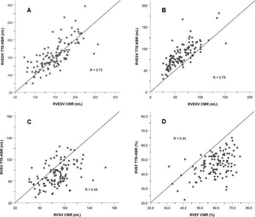
Figure 1. Correlation plots
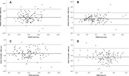
Figure 2. Bland-Altman plots

