


A 61-year old woman presented with progressive dyspnoea, oedema of the lower extremities and a right parasternal diastolic murmur. Echocardiography demonstrated a partly thrombosed, unruptured, non-coronary sinus of Valsalva aneurysm that mimicked a cardiac tumour, resulting in severe aortic insufficiency (Fig. 1 , Videos 1 and 2 ). Computed tomographic angiography confirmed the diagnosis and she underwent successful surgical repair (Fig. 2 ).
Video 1: Live image of parasternal long-axis view of transthoracic echocardiography demonstrated a mass inside the left atrium. The cardiac mass was closely adjacent to the aortic root with an irregular border, which raised the suspicion of a thrombosed sinus of Valsalva aneurysm. Colour Doppler image showed significant aortic insufficiency; the mass also resulted in turbulent and reverse flow inside the left atrium, without interfering with the normal motion of the mitral valve.
Video 2: Live image of parasternal short-axis view of transthoracic echocardiography depicted the huge, non-coronary sinus of Valsalva aneurysm with an intraluminal thrombus and compression of the left atrium. No abnormal blood flow was detected between the aneurysm and the cardiac chambers, and thus there was no evidence of communication between the cardiac chambers and the aneurysm.
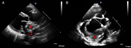
( A ) Transthoracic echocardiography initially showed a huge mass (asterisk) inside the left atrium in the parasternal long-axis view. The border between the mass and the aortic root was uneven, and there was significant aortic insufficiency. ( B ) A short-axis view at the aortic root level eventually revealed that the ‘intracardiac mass’ was an aneurysm with an intraluminal thrombus (asterisk) arising from the non-coronary sinus of Valsalva, with a significant asymmetrically elongated annulus. LA: left atrium.
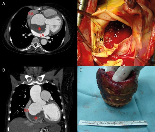
( A and B ) Computed tomographic angiography showed the 8 cm, non-coronary sinus of Valsalva aneurysm with an intraluminal thrombus (asterisks) and externally compressed left and right atria. Significant aortic insufficiency and the aneurysm's external compression of both atria contributed to the clinical manifestations of the patient. ( C ) After cardiopulmonary bypass and an aortotomy, a severely deformed aortic root geometry caused by the elongated annulus of the non-coronary cusp was noted, which made valve-sparing root replacement unfeasible. ( D ) A well-formed, laminated thrombus was found inside the aneurysm and was removed completely. The patient underwent successful composite aortic root replacement plus a porcine bioprosthetic valve implantation with reimplantation of both coronary buttons. RA: right atrium; LA: left atrium; A: aneurysm; L: left coronary cusp; R: right coronary cusp; N: non-coronary cusp.
