


Abstract
Surgical bullectomy is the treatment of choice for giant emphysematous bullae; however, when a giant emphysematous bulla occupies the entire hemithorax, and the remaining lung is in a collapsed state for a long period, it is difficult to predict the surgical outcome preoperatively. We report a case of the successful resection of a giant emphysematous bulla occupying the entire hemithorax. A 44-year old man presented with bilateral giant emphysematous bullae. The giant emphysematous bulla on the left side occupied the entire left hemithorax. Further investigation of past chest radiographs helped predict that the left lung could be re-expanded with the recovery of pulmonary function after resection of the giant emphysematous bulla.
A giant emphysematous bulla is defined as a large bulla occupying at least one-third of a hemithorax [1]. It is difficult to estimate the natural history of these bullae accurately, but their enlargement causes worsening dyspnoea. In patients with a giant emphysematous bulla occupying at least half the volume of a hemithorax, surgical bullectomy is the treatment of choice [2]. However, when the giant emphysematous bulla occupies the entire hemithorax, and the remaining lung is in a collapsed state for a long period, it is difficult to predict the surgical outcome preoperatively. We present a case of giant emphysematous bulla occupying the entire hemithorax; in this case, the left lung could be re-expanded with the recovery of pulmonary function after surgical resection of giant emphysematous bulla.
CASE REPORT
A 44-year old man (chronic heavy smoker, 48 pack-years) was referred to our clinic due to dyspnoea on exertion. Chest radiography showed bilateral giant emphysematous bullae (Fig. 1). Chest computed tomography (CT) revealed that the left giant emphysematous bulla occupied almost the entire left hemithorax. The remaining part of the left lung was small and was compressed to the mediastinum. Pulmonary function tests revealed the following: FVC, 1.64 l (34.3% of predicted); FEV1, 1.03 l (25.1% of predicted); FEV1/FVC ratio, 62.8%; total lung capacity, 3.26 l (42.9% of predicted); residual volume, 1.57 l (74.1% of predicted); diffusion capacity of the lung for carbon monoxide (DLCO), 15.3 ml/mmHg/min (50% of predicted) and DLCO/alveolar volume (VA), 5.64 ml/mmHg/min/l (110% of predicted). Lung perfusion scintigraphy showed that the right-to-left ratio was 97:3. According to chest radiographs taken annually for the past 4 years, the giant emphysematous bulla had already occupied almost the entire hemithorax 4 years previously (Fig. 2), and it had gradually compressed the remaining left lung over time. It was difficult to estimate the cause of the dyspnoea and was also difficult to distinguish giant emphysematous bulla compression of normal parenchyma from the enlargement of an emphysematous lung, but the almost normal DLCO/VA suggested that the remaining lung was not emphysematous. In addition, sequential changes in compression by the giant emphysematous bulla on the annual chest radiographs suggested that the remaining left lung would expand if the compression of the giant emphysematous bulla was removed, and that surgical treatment would relieve dyspnoea felt by the patient upon exertion. Unfortunately, the patient refused surgery on numerous occasions. He presented again 6 months later with a 1-month history of increasing shortness of breath and decreasing exercise tolerance. Chest CT indicated that the left giant emphysematous bulla had grown to occupy the entire left hemithorax with complete atelectasis of the remaining left lung. The patient finally consented to a left bullectomy by video-assisted thoracoscopic surgery. A left-sided, three-port technique was used to gain access to the thoracic cavity. The bullae were not collapsed despite differential ventilation because of air-trapping, and so the walls of the bullae were punctured. The bullae consisted of multiple bullae from the upper lobe fused with a small bulla from the lower lobe. The left lower lobe presented with complete atelectasis. Bullectomy was performed using an endoscopic stapling device (EndoGIA 45 and 60, United States Surgical, Tyco Healthcare, USA) with a synthetic polymer buttress for staple line reinforcement. The staple line was set in the healthy lung tissue along the bases of the bullae. The collapsed left lung was gradually re-expanded by manual ventilation without difficulty. One chest tube was inserted and the left lower lobe was re-expanded without any air leakage. The patient made an uneventful recovery and was discharged. He remained well at the last follow-up 1 year after surgery. Lung perfusion scintigraphy showed that the right-to-left ratio was 62:38. A pulmonary function test revealed the following: FVC, 2.98 l (62.6% of predicted); FEV1, 2.06 l (50.1% of predicted); FEV1/FVC ratio, 69.1%; DLCO, 16.2 ml/mmHg/min (53.2% of predicted) and DLCO/VA, 5.11 ml/mmHg/min/l (83% of predicted).
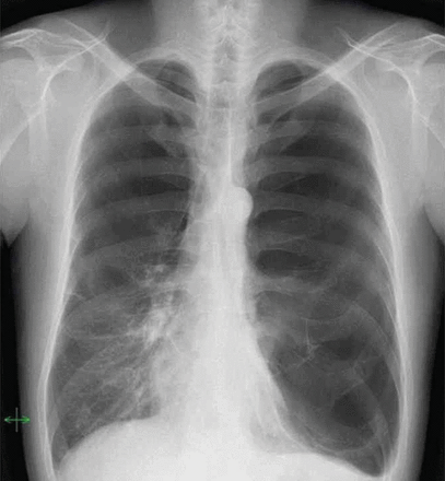
Chest radiography revealed bilateral giant emphysematous bullae. In particular, the left giant emphysematous bulla occupied the entire left hemithorax.
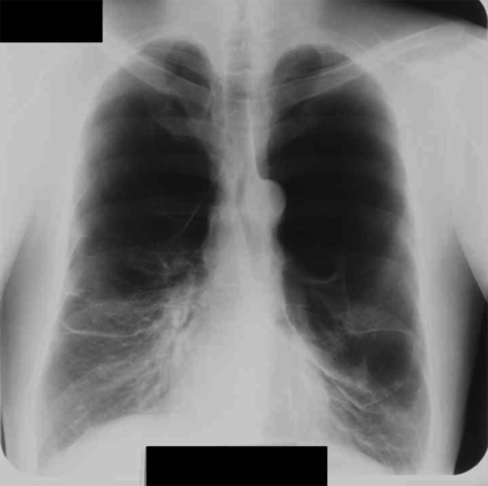
Chest radiograph taken 4 years previously showed that the left lung was compressed by a giant emphysematous bulla, but it was still recognizable.
COMMENT
The natural history of a giant emphysematous bulla without complications is generally characterized by gradual enlargement over time, although spontaneous regression of a bulla is occasionally observed along with the resolution of airway pathology such as an infection, mucus plugging or tumour [3]. Hence, in patients with a giant emphysematous bulla occupying at least half the volume of a hemithorax, surgical bullectomy is the treatment of choice to allow the compressed lung to re-expand with a subsequent improvement of symptoms and lung function [2]. In this case, surgical intervention was the only alternative since the patient had a very large, progressively expanding bulla that was causing compression of the adjacent lung and associated progressive dyspnoea. However, at first, it was difficult to determine whether the giant emphysematous bulla had compressed the underlying normal lung tissue or if the compressed lung itself had become emphysematous and had decreased in size; therefore, the most critical point was to determine whether the collapsed lung could re-expand with sufficient lung function after the bullectomy. There were several pointers supporting the conclusion that the collapsed lung could re-expand. First, almost normal DLCO/VA indicated that the remaining lungs seemed not emphysematous. Secondly, and more importantly, we had obtained past chest radiographs to confirm that the left lung was less compressed and more expanded 4 years ago. On the basis of these data, we performed the bullectomy knowing postoperatively that the left lung could be re-expanded. On the other hand, complete re-expansion and recovery of pulmonary function, despite a long period of complete collapse, has been described elsewhere [4, 5]. It was reported that a selective manual inflation test was useful to assess preoperatively the re-expansion and vascularity of a collapsed lung [5], but our assessment seemed more practical and non-invasive. Furthermore, it might be difficult to perform preoperatively an invasive preoperative study.
In conclusion, we report a case of a giant emphysematous bulla with successful resection. Judging from his normal DLCO/VA and from sequential changes in giant emphysematous bulla on chest radiographs, we preoperatively estimated that the collapsed left lung had almost normal parenchyma and could be re-expanded. Therefore, we presumed that surgical treatment could relieve dyspnoea felt by the patient upon exertion.
Conflict of interest: none declared.
REFERENCES
Author notes
These authors contributed equally to this work.
