


Abstract
Internal mammary arteries graft failure in coronary artery bypass grafting (CABG) is mostly considered to be a result of competitive flow (CF) from the native coronary artery, which significantly limits future revascularization options.
With the left internal mammary artery (LIMA) anastomosed to the left anterior descending (LAD) coronary artery using an off-pump technique, CABG was performed on 15 Chinese swine. Then we produced varying degrees of stenosis in the proximal LAD coronary artery with an adjustable flow occluder, measured the mean flow of the LIMA and LAD coronary artery and detected the plasma concentrations of nitric oxide (NO) and endothelin (ET) in the LIMA graft.
When stenosis of the proximal LAD artery was decreased, the CF of the native LAD coronary artery showed an increasing trend, while the mean flow of the LIMA revealed a decreasing trend. Distal LAD coronary artery flow remained nearly constant, despite the varying proximal LAD artery or LIMA flow rates. An oscillating flow pattern (retrograde/antegrade) was noted in the LIMA graft when the proximal LAD coronary artery was not fully occluded. The plasma concentration of ET in the LIMA graft was significantly higher than that before grafting (P < 0.05), and the plasma concentration of NO was significantly lower (P < 0.05). The concentration of NO was positively related to the mean flow in the LIMA (r = 0.872, P < 0.05).
CF from the native LAD coronary artery can decrease the blood flow in the LIMA graft and even change the direction of the flow. These changes may impair the function of the LIMA graft and affect ET or NO production, which in turn may lead to early LIMA graft failure.
INTRODUCTION
Compared with vein grafts, arterial bypass conduits have a superior long-term patency rate. This is due to the physiological, anatomical, and haemodynamic characteristics of the arterial wall. Accordingly, the use of arterial conduit is now unanimously accepted as the best choice for surgical revascularization [1–3]. However, factors other than arteriosclerosis influence graft patency. An important factor affecting early arterial graft patency is native coronary artery blood flow, even though arteries are muscular and can autoregulate their lumen in response to blood flow demand. Despite the overall excellent patency rates, some have noted increasing frequency of diffuse or distal narrowing of internal mammary artery (IMA) grafts, ultimately leading to graft failure, which significantly complicates future revascularization. Various causes for this entity have been suggested, including damage during harvesting and mobilization, spasm, inflammation as part of a post-pericardiotomy syndrome or a steal phenomenon arising from a large undivided proximal branch of the IMA [4–6]. Moreover, studies have suggested that competitive flow (CF) is also an important factor in early IMA graft failure. Flow competition from minimally diseased native coronary vessels has been implicated in the failure of IMA grafts, but it was not thought to affect saphenous vein graft patency. Few studies have established an association between different degrees of proximal coronary stenosis and graft patency; however, clinical studies of CF do not measure flows because they are retrospective and based on angiographic data [7–9]. In this study, we established a swine model of coronary artery bypass grafting (CABG) with a left internal mammary artery (LIMA) graft to the left anterior descending (LAD) coronary artery, with varying degrees of stenosis in the proximal LAD produced by a flow occluder in order to investigate the influence of CF on LIMA graft flow and the plasma concentration of nitric oxide (NO) and endothelin (ET) in the graft.
MATERIALS AND METHODS
All animals received care in compliance with the Principles of Laboratory Animal Care formulated by the National Society for Medical Research and the Guide for the Care and Use of Laboratory Animals published by the National Institutes of Health (NIH publication 85–23, revised 1996). The experimental animals were 15 Chinese swine weighing 30–45 kg (mean weight, 35.5 ± 3.69 kg). After intravenous pentobarbital induction of anaesthesia (20 mg kg−1), the animals were intubated with a tracheal cannula and ventilated with a volume ventilator. Arterial blood pressure was monitored with a high-fidelity catheter micro-manometer in the left femoral artery. Arterial blood gases were monitored every 30 min, and sodium bicarbonate was added to maintain pH between 7.35 and 7.45. In addition, we ensured that an appropriate anaesthesia depth, continuous oxygen supply and stable blood pressure and heart rates were maintained in order to avoid arterial spasms and ventricular arrhythmias. The electrocardiogram was also continuously monitored.
After trans-sternal thoracotomy under sterile conditions, the LIMA was dissected from the chest wall with a wide pedicle including the accompanying veins and adjacent muscle. The dissection was continued proximally to the vessel's origin at the left subclavian artery and distally to the level of the seventh or eighth intercostal space. Internal mammary arteries were harvested carefully and left covered in papaverine-soaked gauze (30 mg diluted in 30 ml of saline). Blood flow through the LIMA was measured using transit-time flow measurement flow probes (Butterfly flowmeter, Medi-Stim AS, Norway and TS420 flowmeter, Transonic Systems Inc., USA), with both ends of the LIMA still intact in the circulation. The distal end of the LIMA was divided, and blood samples were taken for blood gas analysis and measurements of the plasma concentration of ET and NO as a control.
The pericardium was then incised and its edges were sutured to the margins of the thoracotomy to create a cradle for the heart. The LAD was dissected from the surrounding tissue approximately 1.5 cm proximal to the first diagonal branch (D1) in order to place the occluder and the flow probe, respectively. Coronary shunt, gentle surgery, lidocaine and papaverine was requested during the operation. Topically applied papaverine was used to prevent vasospasm, and when necessary 1% lidocaine was then intravenously administered at a dose of 1.5 mg kg−1 to prevent ventricular arrhythmias. The LAD–LIMA anastomotic site was located between the D1 and the second diagonal branch (D2). The range of LAD diameter was 1.75–2.0 mm, and the average size of the LIMA was ∼2.0 mm. With the help of heart-tissue stabilizer (Octopus II, Medtronic) for heart stabilization and the flow shunt, the anastomosis was constructed using a continuous 7-0 Prolene suture on the beating heart. Coronary intraluminal shunts (1.5–2.0 mm diameter, 10-mm length shunts were selected upon sizing of the vessels) aim to achieve haemostasis at the arteriotomy site and to allow antegrade blood flow using the standard off-pump coronary artery bypass technique. All hearts were allowed to recover for approximately 20 min after the anastomosis, during which time cardiac function became normalized. No evidence of ischaemia was found on the electrocardiogram. An adjustable occluder was placed around the LAD proximal to D1. Three flow probes were used: around the LAD, one probe was placed between the flow occluder and the anastomotic stoma, the other distal to the site of the anastomosis and the third around the LIMA (Fig. 1a and b). In experimental process, minimally invasive surgical techniques, papaverine or lidocaine were used to ensure flow and to avoid arterial spasm and endothelial injury in preparation of arterial conduits (IMA and LAD).
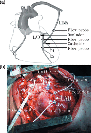
(a) A schematic layout that demonstrates the position of the three flow probes on LIMA and LAD, the standard occluder on LAD and the catheter through LIMA. (b) A real photograph of the experimental swine model of CABG.
The LIMA was initially fully occluded, and the flow in the proximal LAD was measured. After 20 min of equilibration, with both the LAD and the LIMA patent, flows in the LAD proximal and distal to the anastomotic stoma and in the LIMA were measured. Then various degrees of LAD stenosis (30, 50, 75, 90 and 100%) were made with the adjustable occluder by comparing with the flow of the LAD in fully patent when LIMA was occluded to calculate the degree of stenosis. The occluder was adjusted and flow probes were calibrated electronically, confirming adequate haemodynamic parameters and the degree of stenosis. Afterward, blood flows were firstly recorded using the transient transonic flowmeter, and then these data were compared with native flow to calculate the degree of stenosis. The fine-needle catheter placed into the LIMA distal branches by puncture was used to collect blood samples for testing. IMA graft flow distal to the catheterized site was measured using the flow probe and compared with native IMA flow to confirm catheter has little effect on blood flow in experimental process. Topical papaverine was used on the catheter site to prevent arterial spasm and sequentially measured blood flow to ensure that a baseline level had been reached. We recorded the mean flows at each probe and took blood samples from the distal end of the LIMA with a catheter (Fig. 1a and b) after 5–10 min of equilibrium at each occlusion level, respectively. Before and after constructing anastomosis, and with or without CF, all flow measurements were made after 10 min stabilization and at similar and stable haemodynamic conditions (heart rate and blood pressure). The plasma concentration of ET was measured by the radioimmunoassay method and that of NO by nitrate reductase method. Both kits were made by Nanjing Jiancheng Bioengineering Institute.
Statistical analysis
Results were statistically analysed by one-way analysis of variance and Pearson relating with SPSS for Windows 12.0 software (SPSS Inc., Chicago, USA). A P-value <0.05 was considered significant.
RESULTS
Mean flow of the left internal mammary artery graft
We observed that the higher the degree of stenosis in the proximal LAD, the lower the CF in the LAD and higher the mean flow in LIMA. The mean flows of the LIMA at the degrees of 0, 30 or 50% were significantly lower than that of the LIMA at the degrees of 90 or 100% (P < 0.01). Meanwhile, no statistically significant difference was found in LAD flow distal to the site of the anastomosis at different degrees of stenosis (P > 0.05, Fig. 2).
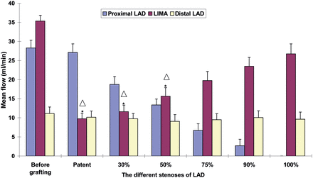
The mean flows of LIMA and LAD proximal and distal to the anastomosis. *P < 0.01 vs 90% stenosis; △P < 0.01 vs 100% stenosis.
Flow waveform of left internal mammary artery graft
We classified the LIMA flow into diastolic flow and systolic flow. The direction of the diastolic flow of LIMA was forward whether the LAD was occluded or not. The systolic flow of LIMA was lower than the diastolic flow. Furthermore, oscillating flow (retrograde/antegrade) waveforms were observed in 15 swine at 0, 30 and 50% stenosis of the LAD, in 11 at 75%, in 6 at 90% and in 0 at 100%. Typical biphasic velocity waveforms were shown in Fig. 3.
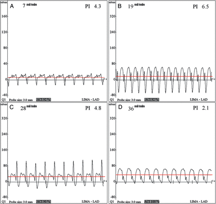
LIMA flow waveforms with the proximal LAD at four different degrees of stenosis: 0 (A); 50 (B); 75 (C); and 100% (D). Typical biphasic velocity waveforms were noted at 0 (patent) and 50% stenosis. The upper lines represent the mean flow of LIMA.
Plasma concentration of NO and ET
The plasma concentration of NO in LIMA after grafting was significantly lower than that before grafting (P < 0.05, Fig. 5). The concentration of NO in the LIMA graft was significantly lower when the stenosis of the proximal LAD was 0, 30 and 50% than that when the stenosis was 90 or 100% (P < 0.05). The concentration of NO was positively related to the mean flow of the LIMA graft (r = 0.872, P < 0.05, Fig. 4). The plasma concentration of the ET in LIMA after grafting was significantly higher than that before grafting; however, there was no statistical significance between different degrees of stenosis after grafting (Fig. 5).
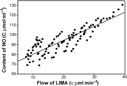
A scatterplot demonstrates the relation between LIMA flow and concentration of NO. The content of NO was positively related to the mean flow of the LIMA graft (r = 0.872, P < 0.05) and oblique line represents the line of regression equation.
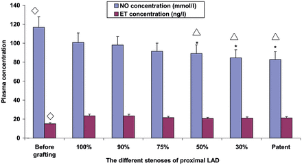
The mean concentration of NO and ET in LIMA at different stenosis. *P < 0.05 vs 90% stenosis; △P < 0.05 vs 100% stenosis; ⋄P < 0.05 vs after grafting.
DISCUSSION
Vein graft patency was not satisfactory; it declined with time from development of graft arteriosclerosis. Because of their long-term patency and their freedom from atherosclerosis, IMAs have been widely used as the most reliable graft material in prolonging patient survival and preventing recurrent ischaemic events. There are, however, increasing reports of graft failure due to diffuse or distal narrowing. The spasm and inflammation of the IMA graft, steal from the proximal branches and damage to the IMA during mobilization and anastomosis may account for stenosis [10–12].
There is a considerable body of clinical and experimental evidence to suggest that endothelial signals may modulate downsizing of the vessel diameter under CF conditions. Some studies also hypothesize that CF from the native coronary arteries, to which the IMAs were anastomosed, is the cause of failure [13–15]. Villareal and Mathur [16] prospectively reviewed 261 patients who had undergone IMA grafting and found 28 cases of IMA graft failure and 22 of them were under competitive-flow conditions, whose angiography before surgery showed that the recipient vessels had less-than-critical stenosis (<70%). Moreover, the study of Berger and colleagues indicated that the occlusion rate of IMA was very high (79%) when the stenosis of proximal LAD was <50% [17]. These results suggested that the degree of stenosis in the native vessel and the CF influenced the patency of the IMA bypass graft.
But some studies demonstrated that even under relatively low flow conditions, the IMA may retain its auto-regulatory capacity, at least in the short-term. Spence et al. [18] and Lust et al. [19], using a canine model of CF, showed that at least acute IMA graft flow is maintained above in situ levels even when grafted to a completely patent coronary artery. Even after 2 months of maximal chronic flow competition from a fully patent native artery, IMA graft flow was maintained above in situ levels, and a recruitable flow reserve from the IMA graft could be demonstrated when the native vessel was occluded. Their available data leave considerable doubt about the CF mechanism of mammary artery narrowing and failure.
However, we must note that these competition flow studies described IMA anastomosed to the left circumflex artery (LCX) in a canine model, wherein ischaemic tolerance of heart, structure and function of coronary artery may be different from that in pigs. In addition, the angle between LCX branch and left main coronary artery is ∼90° and the relative blood flow in LCXs is much less than that in LADs. But ischaemic sensitivity or arterial spasms in pigs are similar to that in humans, and the LIMA anastomosed to the LAD is most clinically common. Therefore, the impact of CF may be different with IMA grafted to the different branch of the coronary artery.
Furthermore, some researches revealed that a narrowing phenomenon of the mammary graft was associated with CF and this phenomenon was identical with other previously results. Nordgaard et al.'s [7] study showed that the LIMA graft was much influenced by CF, as both the full and partial CF conditions significantly altered the flow patterns. Other studies [16, 17] showed that internal thoracic artery patency decreases as coronary CF increases and reduction of CF results in increased distal native coronary flow.
Although CF was considered as a tenable theory of graft stenosis, the mechanism has not been elucidated. Some studies found that reduced IMA flow resulted from high flow rates in the native LAD damaged the endothelial cells and ultimately led to IMA shrinkage [16, 17]. Modern data have pointed out the importance of a functionally preserved endothelial layer in the arterial conduit. Endothelial cells can secrete endothelium-derived relaxing factors (EDRF) and endothelium-derived contracting factors (EDCF), whose content was maintained in a dynamic balance in a normal physiological environment. NO, the most important EDRF that can relax blood vessel walls, restrains the proliferation of vascular smooth muscle cell (VSMC), restrains the aggregation and adhesive attraction of platelet and prevents vasospasm and thrombogenesis. On the other hand, ET is the most important EDCF that can cause vasoconstriction and promote the proliferation of VSMC. Therefore, we hypothesize that CF may result in IMA stenosis through the impairment of the endothelial cells changing the balance of NO and ET. If the endothelium is damaged, there may be a strong spasmodic effect because of the loss of EDRF, which appears to be due to the preservation of intrinsic vasodilatation mechanisms [20–23]. Besides, we all know that the non-muscular vein grafts cannot adjust their lumens in response to metabolic requirements as much as arterial grafts, but vasospasm is one of the most important factors influencing blood flow in the artery graft model. Vasospasm may be caused by multiple mechanisms and different causes, and more attention should be paid to this problem. Mechanical stimulation by the surgical devices on the IMA and LAD artery wall is frequently responsible for the vasospasm. Some drugs (lidocaine and papaverine) have been used to reduce spasm and augment flow following vascular bypass procedures. Papaverine, which induces vasodilatation by direct action on smooth muscle, was often used to treat arterial spasm. Topical lidocaine and papaverine can be used in the preparation of IMA and LAD, because they can increase the blood flow, prevent arteries spasm and may increase the diameter of arteries [23–25]. The conduit should be handled carefully to prevent endothelial injury and consideration should be given to dilatation by mechanical and pharmacologic means.
Our study aimed to assess the impairment of graft flows due to CF from the coronary artery or stenosis of the coronary anastomosis. Transit-time flow patterns were measured at different flow conditions in the LIMA graft to LAD of a porcine model. The data of our study showed that the higher the degree of stenosis in the proximal LAD, the lower the CF, which was in accord with previous studies. The mean flows of the LIMA at 0, 30 or 50% LAD stenosis was significantly lower than that of the LIMA at 90 or 100% stenosis, which indicated that the degree of stenosis in LAD resulted in the generation of CF that had important effects on IMA graft flow. This study showed that the LIMA graft was much influenced by CF, as both the full and the partial CF conditions significantly altered the flow patterns. Our results also found that the higher the CF of the proximal LAD was, the lower the plasma concentration of NO in the LIMA graft. The concentration of NO was positively related to the mean flow in the LIMA graft (r = 0.872, P < 0.05). Furthermore, in our experiment, an oscillating flow (retrograde/antegrade) was noted in the LIMA when the proximal LAD was not fully occluded. We suspect that the low and even oscillating flow may damage the endothelial cells of the LIMA decreasing the concentration of NO, resulting in a breakdown of the dynamic balance between ECRF and EDCF, ultimately leading to IMA stenosis. On the basis of the data of our study, CF from the proximal LAD has important effects on the IMA graft flow and the concentration of NO and ET, predisposing the IMA grafts to stenosis.
In our model, the LIMA flow was significantly reduced by native coronary artery CF, and CF changes have stimuli to the arterial wall endothelium, with the release of NO and ET. This may injure the endothelium or alter endothelial-mediated signals, and endothelial signals may regulate the vessel diameter when faced with these NO and ET stimuli, that may be involved in the development of arterial graft failure.
This is only an initiatory study. The limitations of this study were that the pigs had normal coronary arteries and this animal model may not be directly comparable to patients with advanced coronary disease. Some factors just like vascular resistance, vasospasm and adaptability may play an important role in the changes of bypass flow after operation. With these drawbacks, results from CF studies involving IMA graft and coronary artery should be correlated to the human setting very cautiously. The observational data from this study, however, allowed us to postulate the endothelial mechanism of CF. The true reason and explicit mechanism of IMA failure are still not well known. Detailed studies are needed in the future.
ACKNOWLEDGEMENTS
We gratefully acknowledge the advice from Henry Low and Christopher Clyne in Hartford Hospital.
Conflict of interest: none declared.
