


Abstract
Dynamic performance of the aortic valve (AV) after ascending aorta replacement with reimplantation of the native AV (David) was investigated.
We prospectively evaluated 17 patients who underwent David procedure. Rest/stress echocardiography follow-up was performed and results were compared with those of matched healthy controls.
There were no significant differences in terms of age, height, weight, BSA, left ventricular mass, left ventricular ejection fraction (LVEF) and tele-diastolic volume between the David and control group. At rest echocardiography, patients in the David group had a lower indexed aortic valve area (IAVA) (1.1 ± 0.2 vs. 1.5 ± 0.2 cm2/m2, P < 0.0001), with comparable transvalvular gradients (TVG). At maximal physical stress, although the IAVA in the David group was significantly increased from the rest values (P = 0.001), the difference with the control group persisted (David 1.4 ± 0.3 vs. Control 1.7 ± 0.2 cm2/m2, P < 0.0001) maintaining similar peak TVG (David 13.6 ± 5.3 vs. Control 11.7 ± 4.5 mmHg, P = ns) and mean TVG (David 7.2 ± 3.0 vs. 6.2 ± 2.4 mmHg, P = ns). AV regurgitation in the David group was absent in five (29.4%), grade I in nine (52.9%) and grade II in three (17.6%) patients and remained unchanged during stress. At multiple linear regression, David operation was inversely correlated to rest IAVA (OR = −0.4; P = 0.01; CI: −0.7–0.1).
Although IAVA is significantly smaller after David procedure in comparison with matched controls, no pathological increase in TVG is noticed. A significant increase in the IAVA during physical stress documents the preserved pliability/elasticity of the aortic unit after David procedure preventing pathological increase in the TVG even during strenuous effort.
INTRODUCTION
Description of valve sparing procedures by Yacoub in 1979 [1] and David in 1988 [2], respectively, has sparked tremendous interest in reparative techniques of the aortic valve (AV), somehow neglected up to that time in comparison with the mitral valve. As a result, the anatomy and physiology and the importance of the aortic root complex as a single unit have been clarified in detail. Despite the outstanding results reported in the literature [3, 4], however, the fate of the AV complex after remodelling or reimplantation procedures remains an object of much debate [5]. On the other hand, exercise echocardiography has become an important tool in revealing the dynamics of the valve and the ventricle and unmasking functional disabilities in patients who often adapt by reducing their physical activity [6]. In the light of these considerations we became interested in studying by means of echocardiography the behaviour of the AV complex after a reimplantation procedure, at rest and after exercise, in comparison with normal subjects.
MATERIALS AND METHODS
Twenty-one consecutive patients underwent a reimplantation procedure (David I) between 2004 and 2009. All patients had anatomically normally shaped tricuspid AVs free from any sort of leaflet alteration. The diameter of the sinotubular junction determined the diameter of the graft, with an additional over-sizing of 1–2 mm. Straight Dacron grafts, sized 26, 28 and 30, were used.
One patient died of gastric cancer 1 year after the procedure and three patients were lost to follow-up. Seventeen patients were available for study. For every patient previously submitted to the David procedure, an adequate match for gender, age, body surface area (BSA) and LVEF% was found within the group of patients who are daily referred to our echocardiography clinic for a medical check-up. Patients in the two groups (David and Control), for this reason, belonged to the same referral area. A group of 18 subjects was identified for matching and comparison. The two groups were studied at baseline and after treadmill exercise according to Bruce's protocol with the targets of 75% of predicted maximal heart rate or appearance of symptoms [7].
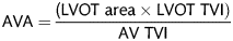
Measurements of left ventricular (LV) dimensions were made from 2D echocardiographic images in the parasternal long-axis view and M mode. LV volumes and ejection fraction (EF%) were calculated by modification of Simpson's method with two apical views. AV regurgitation was graded on the basis of the regurgitant jet height/left ventricular outflow tract (LVOT) height ratio (mild (1+): <25%, moderate (2+): 25–46% , moderately severe (3+): 47–64% and severe (4+): ≥65%).
All patients had signed a written consent to the collection and eventual analysis of their personal medical data.
STATISTICAL ANALYSIS
Data were expressed as means ± standard deviation. Comparison between the groups (David and Control) was performed using the Student's t-test for independent samples after having tested the normal distribution of the different variables. Normality of continuous variables was tested by means of the Wilk–Shapiro test. AVAs in the David group at rest and at maximal stress were compared using the paired Student's t-test. Multiple linear regression was performed to identify the correlation between rest AVA (dependent variable) and group characteristics. SPSS 14.0 was used to perform the statistical analysis.
RESULTS
All continuous variables investigated were normally distributed. There were no significant differences in terms of age, height, weight, BSA, LVM, LVEF and tele-diastolic volume between the David and control group (Table 1). AV regurgitation in the David group was absent in five (29.4%), grade I in nine (52.9%) and grade II in three (17.6%) patients. At rest echocardiography, patients in the David group had a significantly lower-indexed AVA (IAVA) (David 1.1 ± 0.2 vs. Control 1.5 ± 0.2 cm2/m2, P < 0.0001), with comparable transvalvular gradients (TVG) (Table 2). At maximal physical stress, although the IAVA in the David group was significantly increased from the rest values (rest AVA 1.1 ± 0.2 vs. peak stress AVA 1.4 ± 0.3; P = 0.001), the AVA difference between the groups persisted maintaining similar transvalvular peak and mean gradients (Table 2). Mean AV regurgitation in the David group remained unchanged during stress. Because AVA was the only echocardiographic variable differentiating the David from the control group, we tried to identify the correlation between rest AVA (dependent variable) and characteristics within the entire study cohort. At linear regression analysis, rest AVA was inversely related to the David procedure only (Table 3). Although LVM seemed to be directly related to AVA (OR = 0.3; P = 0.04; CI 95%: 0.0–0.005), the OR value did not fall within the confidence interval (Table 3). No patient experienced cardiac symptoms during the testing.
Baseline characteristics in the two groups: David and Control
| Mean | SD | ||
|---|---|---|---|
| Age | Control | 60.2 | 8.0 |
| David | 64.7 | 7.6 | |
| Height (cm) | Control | 167.2 | 9.7 |
| David | 168.1 | 6.6 | |
| Weight (kg) | Control | 76.3 | 11.9 |
| David | 82.1 | 18.7 | |
| BSA (m2) | Control | 1.85 | 0.1 |
| David | 1.90 | 0.2 | |
| LVM (g) | Control | 241.4 | 77.1 |
| David | 209.1 | 48.0 | |
| LVEF % | Control | 56.0 | 7.1 |
| David | 52.5 | 9.4 | |
| TDV (ml) | Control | 102.2 | 24.3 |
| David | 109.2 | 35.7 |
Baseline and peak stress echocardiography data
| Mean | Standard deviation | P value | ||
|---|---|---|---|---|
| B_Aortic max velocity (cm/s) | Control | 134.1 | 24.1 | 0.4 |
| David | 140.9 | 29.6 | ||
| B_Aortic mean velocity (cm/s) | Control | 95.1 | 18.5 | 0.9 |
| David | 94.5 | 24.1 | ||
| B_Aortic Mx grad (mmHg) | Control | 7.3 | 2.6 | 0.3 |
| David | 8.3 | 3.8 | ||
| B_Aortic mean grad (mmHg) | Control | 4.1 | 1.6 | 0.7 |
| David | 4.3 | 2.1 | ||
| B_AVA (cm2) | Control | 2.8 | 0.4 | 0.000 |
| David | 2.2 | 0.3 | ||
| B_AVA indexed (cm2/m2) | Control | 1.5 | 0.2 | 0.000 |
| David | *1.1 | 0.2 | ||
| P_Aortic max velocity (cm/s) | Control | 168.7 | 33.2 | 0.2 |
| David | 182.5 | 36.1 | ||
| P_Aortic mean velocity (cm/s) | Control | 116.2 | 23.7 | 0.5 |
| David | 122.1 | 28.4 | ||
| P_Aortic Mx grad (mmHg) | Control | 11.7 | 4.5 | 0.2 |
| David | 13.6 | 5.3 | ||
| P_Aortic mean grad (mmHg) | Control | 6.2 | 2.4 | 0.3 |
| David | 7.2 | 3.09 | ||
| P_AVA (cm2) | Control | 3.2 | 0.5 | 0.002 |
| David | 2.6 | 0.5 | ||
| P_AVA indexed (cm2/m2) | Control | 1.7 | 0.2 | 0.002 |
| David | *1.4 | 0.3 |
B: Baseline; P: peak stress; AVA: aortic valve area. Notice that at maximal physical stress, the IAVA in the David group is significantly increased from the rest values (*P = 0.001).
Linear regression analysis
| Beta (OR) | P value | CI 95% | ||
|---|---|---|---|---|
| LL | UL | |||
| David | −0.4 | 0.01 | −0.74 | −0.11 |
| Age | −0.2 | 0.1 | −0.03 | 0.004 |
| Height (cm) | −0.5 | 0.4 | −0.14 | 0.03 |
| Weight (kg) | −1.7 | 0.2 | −0.11 | 0.04 |
| BSA | 2.0 | 0.2 | −4.02 | 13.6 |
| LVM (g) | 0.3 | 0.04 | 0.0 | 0.005 |
Dependent variable: Aortic valve area at rest.
BSA: body surface area; LVM: left ventricular mass; CI: confidence interval; LL: lower limit; UL: upper limit; OR: odds ratio.
COMMENT
To our knowledge, dynamic performance of the AV after ascending aorta replacement with reimplantation of the native AV has never been evaluated. Furthermore, no author has ever compared the performance of reimplanted AV to that of AV belonging to healthy controls. In our opinion, it is essential that the physiologically dynamic performance of the reimplanted AV is evaluated and preserved particularly in young patients prone to physical stress.
Although the group presented in this article is small, all patients were operated upon using exactly the same technique and the stress echocardiography were all performed by a single operator following a very timely protocol.
The study confirmed that the native AV, reimplanted in a straight Dacron tube behaves, after exercise, much like the valve of normal subjects. Although IAVA is significantly smaller after the David procedure, in comparison with matched controls, no pathological increase in TVG is noticed at rest or even during strenuous stress. Furthermore, a significant increase in the basal IAVA during physical stress documents the preserved pliability and elasticity of the aortic unit after the David procedure that prevents a pathological increase in the TVG. The higher baseline gradient and smaller valve area of reimplantation patients probably result from the anuloplasty effect of the straight tube sown directly on the annulus. This is confirmed in our multivariate analysis where the David procedure has the strongest correlation (inverse) with the indexed AVA at rest.
An intense discussion has been reported in the literature over the key role of the sinuses of Valsalva on the closing mechanism of the leaflets and the reduced stress whenever they are preserved [9–13]. Furthermore, distensibility of the prosthetic root seems reduced compared with the native root, owing to the noncompliant prosthetic material leading to an increased bending deformation index of the leaflets. Whether all these considerations will have an impact on long-term durability of the valve remains speculative at this time.
The absence of sinuses in our study group did not seem to have any influence on the parameters studied. It is interesting to observe an apparently normal function of the reimplanted AV in a straight tube as described originally by David, before the technical modifications (Valsalva graft, David IV and V) brought by the physiologic observations of recent years. In our study, fixation of the annulus and of the commissural posts inside a rigid tube does not alter the physiology of the valve. After all, it may very well be proved that, at long-term follow-up, the reimplanted valve works well even in the absence of the sinuses of Valsalva.
LIMITATIONS
The present study includes a limited cohort of patients. Although our statistical findings are significant and are supported by our clinical hypothesis, the effects of the David procedure on the dynamic performance of the AV should be investigated in larger samples. Furthermore, the cohort in analysis was operated upon during a relatively extended time span (∼5 years) and the mean age of the group seems to be relatively high for patients submitted to a David operation. In reality, we submit to this operation only those patients who have an anatomically perfect AV (free from any sign of thickening or initial calcification) and this is reflected in the limited cohort size. Moreover, patients' mean age refers to the value at the time of follow-up (for matching reasons we could not consider age at the time of surgery) and as a result is higher than expected. In any case, we do not have a specific age cut-off for the David operation, as long as the AV has a preserved anatomy. Finally, an analysis of the effects of intraoperative variables and follow-up duration on the haemodynamic performance of the AV was not performed and was beyond the aim of our analysis. In fact, the aim of the study was to determine whether the David procedure could, per se, have an impact on the AV performance at rest and under physical stress.
Conflict of interest: none declared.
