-
PDF
- Split View
-
Views
-
Cite
Cite
Po-Chih Chang, Wen-Lun Wang, Tzer-Zen Hwang, Yu-Jen Cheng, Intramural dissection with mucosal rupture alleviating phlegmonous esophagitis, European Journal of Cardio-Thoracic Surgery, Volume 41, Issue 2, February 2012, Pages 442–444, https://doi.org/10.1016/j.ejcts.2011.06.027
Close - Share Icon Share
Abstract
We report a woman presenting with unrelenting odynophagia and chest pain. Computed tomography identified a deep neck infection with acute phlegmonous esophagitis. However, esophageal intramural dissection with mucosal rupture occurred after routine nasogastric-tube insertion, and pus was vomited thereafter. The patient was treated with antibiotics and delayed endoscopic closure of the rupture site and made a full recovery. Although the definite pathogenesis remained unclear, esophageal intramural dissection with mucosal rupture, a possible and rare complication of nasogastric-tube insertion, eventually alleviated the acute phlegmonous esophagitis in our patient.
INTRODUCTION
The occurrence of ‘esophageal’ intramural dissection is usually due to iatrogenic causes, such as nasogastric (NG)- tube insertion or endoscopy [1,2]. An underlying etiology or abnormal anatomy, such as inflammation, intramural hematoma, stricture, or diverticula may complicate NG-tube insertion and predispose to this intractable condition [2]. However, acute phlegmonous esophagitis, a rare esophageal pathology, has never been reported to be associated with intramural dissection [3,4]. Herein, we report the case of a patient with acute phlegmonous esophagitis secondary to a deep neck infection, in which esophageal intramural dissection occurred after NG-tube insertion.
CASE REPORT
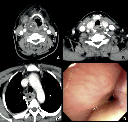
A 57-year-old, well-nourished female presented with a sore throat for 5 days and subsequent odynophagia, dysphagia, and chest pain. Her medical history was remarkable only for hypertension. Physical examination was noteworthy for tachypnea, stridor, and cervical skin inflammation. Computed tomography (CT) was performed because of a high degree of suspicion of deep neck infection and revealed abscesses at the retropharyngeal and right parapharyngeal spaces and diffuse thickening of the esophageal wall (Fig. 1(A)). In addition, a hypodense lesion was seen within the esophageal wall (Fig. 1(B) and (C)). Initially, an esophagogram with a water-soluble contrast medium revealed no evidence of leakage. Only a transnasal ultrathin endoscope (GIF-XP260N; Olympus Optical Co, Ltd., Tokyo, Japan) could be passed, and severe swelling at the arytenoid and upper esophagus with intact mucosa was noted (Fig. 1(D)). No endoscopic ultrasonography could be performed at that time. Urgent bilateral cervicotomy with drainage and tracheostomy were performed due to a concern of sepsis and respiratory distress. At surgery, pus was evacuated from the neck and the superior mediastinum, and the outer esophageal muscle layer was noted to be intact. Because of the potential for esophageal perforation, no concomitant esophageal procedure was conducted. The patient was treated with amoxicillin/clavulanate and metronidazole; an NG tube was placed for nutritional support at that time.

(A) Axial CT of the neck revealed a deep neck infection. (B and C) Chest CT revealed circumferential thickening of the esophageal wall (arrow) with an intramural lower density lesion (arrowhead). (D) Endoscopy revealed diffuse luminal narrowing with poor distensibility at the upper esophagus.
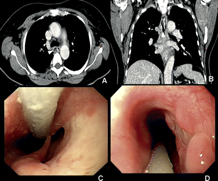
After a changing of the NG tube on postoperative day 10, the patient experienced a sudden onset of retrosternal pain, and copious pus was expelled from the mouth after NG-tube removal. An esophagogram to exclude possible esophageal perforation did not reveal obvious contrast extravasation. The NG tube was carefully reinserted, and the subsequent chest CT revealed an esophageal intramural dissection without obvious mediastinal contamination (Fig. 2(A) and (B)). Esophagogastroendoscopy demonstrated a double lumen with purulent discharge from the dissection orifice (Fig. 2(C)). To drain the phlegmonous infection thoroughly, we performed a delayed endoscopic closure of the dissection orifice with hemoclips on the 35th hospital day. The final culture of the purulent fluid yielded Klebsiella pneumoniae.

(A and B) Axial and coronary chest CT demonstrated an esophageal intramural dissection (arrowhead) after NG tube exchange. (C) Endoscopy revealed an esophageal double lumen, compatible with esophageal intramural dissection. (D) Repeat endoscopy 3 months later showed a well-healed dissection lumen.
After continued administration of broad-spectrum antibiotics and surgical drainage of the deep neck infection twice, the patient recovered well, and interval healing of the esophageal intramural dissection was noted (Fig. 2(D)).
Further, she was able to remain well nourished through NG feeding. The patient resumed oral intake 3 months later without difficulty.
DISCUSSION
A deep neck infection is a serious condition that may ultimately progress to upper-airway compromise, descending necrotizing mediastinitis, or sepsis [5]. However, esophageal involvement is infrequent. Acute phlegmonous esophagitis, an unusual infection with a pattern of mucosal sparing, has been rarely reported to be associated with deep neck infections [3–5]. Although the pathogenesis of acute phlegmonous esophagitis still remains unclear, it is believed that an immunocompromised status such as is associated with either diabetes or alcoholism may predispose patients to this condition [3,4]. However, our patient had been in good health, and no disease such as diabetes or alcoholism was reported. We surmised that purulent fluid descending from the deep neck infection dissected the posterior mediastinum along the retrovisceral space and ultimately infiltrated the esophagus, leading to the phlegmonous infection. CT is the gold standard for a definite diagnosis of acute phlegmonous esophagitis [3,4]. Typical findings are a low-density circumferential area within the thickened wall of the esophagus. Moreover, endoscopic examination and contrast esophagogram are mandatory, so as to exclude inner mucosal lesions and possible esophageal perforation [3,4]. The condition usually responds well to broad-spectrum antibiotics; however, drainage with esophageal myotomy and mediastinal debridement is necessary for concomitant mediastinal involvement [3,4]. Presumably due to its rarity, the use of endoscopic internal drainage for acute phlegmonous esophagitis has not been discussed in the literature.
Early resumption of enteral feeding via an NG tube will facilitate adequate nutrition in patients with deep neck infections [5]. For those with concomitant esophageal involvement, especially esophageal mucosal injury, an alternative solution with feeding gastrostomy or jejunostomy should be executed to avoid devastating complications due to possible esophageal perforation [6]. Though a simple procedure, NG-tube insertion itself may cause undesirable complications, such as upper aerodigestive tract injury or esophageal perforation [2]. Esophageal intramural dissection, which is extremely rare, has seldom been reported with NG-tube insertion [2]. To the best of our knowledge, the concurrent presence of acute phlegmonous esophagitis and esophageal intramural dissection has never been described in the literature. However, this unfavorable complication in our patient after NG-tube exchange, with the function of ‘internal drainage’, led to the evacuation of the phlegmonous infection incidentally. The possible mechanism is that the expanding phlegmonous infection within the esophageal wall made the esophagus prone to intramural dissection, and NG-tube insertion caused a sudden pressure change creating a mucosal injury, which allowed the pus to be expelled [7].
Although esophageal intramural dissection can be managed with conservative methods, such as parenteral nutrition only or a minced diet with proton-pump-inhibitor treatment, the use of endoscopic treatment for esophageal perforation or intramural dissection has been widely accepted in selected patients [1,2,6,8]. So as to evacuate the abscess within the esophageal wall completely and prevent possible mediastinal contamination due to esophageal perforation, we delayed the closure of the dissection orifice, instead of an incision of the dissected septum, and treated this patient successfully with antibiotics.
In conclusion, though the outcome was favorable, we would like to emphasize that an NG tube should not be inserted without caution, when treating patients with underlying esophageal pathologies.
Conflict of interest: none declared.


