


Abstract
We report here an uncommon anterior chest trauma with an unusual fatal penetrating coronary artery injury by pneumatic nail gun with effective perioperative management. While doing upholstery, a 32-year-old male patient accidentally stabbed by a pneumatic nail gun with injury to the anterior chest was brought to the emergency room of our hospital. Persistent chest pain with unstable vital signs and no external injury except for a faint ecchymosis on anterior chest were noted at arrival. Sixty-four-slice computed tomography (CT) scan revealed a foreign body completely embedded in the chest wall penetrating the left ventricle, with the coronary artery also suspected of being involved because of ST-T changes of V2 to V6 on electrocardiography. Three-dimensional reconstructive CT scans showed a penetrating injury to the left anterior descending coronary artery without complete transection. Thereafter, we performed the operation of nail removal with direct repair of coronary artery that was scheduled based on the image findings preoperatively, and the operation was smoothly performed without coronary artery cardiopulmonary bypass and grafting bypass effectively and simply. He was discharged uneventfully 14 days later. Another CT scan was performed which showed patency of repaired coronary artery 3 months later.
INTRODUCTION
Mortality from penetrating cardiac injury was much higher in World War I than in World II and has decreased in recent decades [1]. More victims of penetrating cardiac injury survive the initial trauma with improvement in the overall management of injured patients. More injuries with highpower and -speed missiles are occurring now; besides, major coronary artery injury in penetrating cardiac trauma is relatively uncommon and is associated with a poor prognosis. However, while several cases have been reported before describing emergent cardiopulmonary bypass (CPB) and coronary artery bypass grafting (CABG) for penetrating coronary artery injury, we present here a case of incomplete transaction of the left anterior descending (LAD) coronary artery managed by primary repair of vessel guided by the three-dimensional reconstructive computed tomographic (CT) scans of the coronary artery.
CLINICAL SUMMARY
A 32-year-old male was sent to our emergency department for an accidental pneumatic nail gun injury of the anterior chest 90 min after accident. On arrival, his blood pressure was 90/68 mmHg with heart rate of 108 beats/min, respiratory rate 20 breaths/min, and temperature 36.8 °C without apparent venous engorgement of the neck. Furthermore, persistent chest pain without predominant external injury except for a pin-hole-like ecchymosis on anterior chest was noted. The following cardiac enzymes, when arrival, were recorded: creatinine kinase (CK) 1.65 (26–174 IU l−1); creatinine kinase-MB (CK-MB) 19.1 (0.4–6.3 ng ml−1); and Troponin-I 1.65 (<0.04 ng ml−1). This patient had had a traumatic acute coronary syndrome presenting with ST-T elevation of electrocardiography and cardiac enzyme elevation. Subsequent chest X-ray revealed a foreign body in the cardiac shadow without pneumo- or hemo-thorax.
Because of his relative stable presentation and vital signs, we arranged a 64-slice CT scan thereafter, which revealed a foreign body penetrating into the left ventricle, and the LAD coronary artery was suspected of being involved because of ST-T changes of electrocardiography over V2 to V6 (Fig. 1 (A) and (B)). The three-dimensional reconstructive 64-slice CT scan of the coronary artery showed a penetrating injury of the LAD without complete transection (Fig. 1 (C)), with little amount of hemopericardium, and without pneumopericardium.
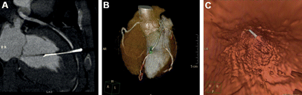
(A) Preoperative 64-slice computed tomography scans revealed a nail penetrating into the left ventricle with suspicion of coronary artery involvement; (B) the nail was totally buried in the chest wall. (C) Three-dimensional reconstruction of the coronary artery (LAD) with walk-through motion pictures (not shown in this figure).
In view of hemodynamic stability, we thought that the incomplete transaction of the LAD could be managed by an effective and simple procedure. Based on the finding of reconstructive CT scans preoperatively, we decided to remove the nail and directly repair the LAD by 6/0 polypropylene sutures under the beating heart without coronary artery CPB (Fig. 2(A)), and the operation was performed 30 min after arrival to our emergent department and standard sternotomy performed with nail removal and direct repair of coronary artery as planning without difficulty.

(A) A nail hooked into the chest wall with coronary artery penetration; 160 ml bloody pericardial effusion was sucked out. (B) and (C) Postoperative 64-slice computed tomography scans with three-dimensional reconstruction of the coronary artery showed no stenosis of LAD.
The patient recovered uneventfully after operation. The following cardiac enzymes were CK 1617 IU l−1; CK-MB 151.4 ng ml−1; Troponin-I 12.53 ng ml−1 just after the operation. Cardiac enzymes reached peak 3 days later and returned back to CK 65 IU l−1; CK-MB 1.1 ng ml−1; and Troponin-I 0.14 ng ml−1 when he was discharged 14 days later.
Another three-dimensional reconstructive CT scan and electrocardiography were performed which showed patency of LAD and a normal electrocardiography 3 months later (Fig. 2(B) and (C)).
DISCUSSION
As per previously published reports, the most common intracardiac foreign bodies are fragments of central venous catheters, bullets, and needles [2]. Penetrating cardiac injuries have high mortality ranging from 12% to 62% [3,4]. The unusual consequence of an untreated penetrating cardiac wound is death. With early diagnosis, rapid transportation, and sufficient resuscitation, mortality can be greatly reduced [5]. However, penetration into the coronary artery in penetrating cardiac trauma is relatively rare (after a review of the literature), and we did not find any report of the retention of a pneumatic nail gun projectile inside the coronary artery [2]. For a variety of reasons, the outcome of these patients has been improving during the last two decades. The main factor is the use of emergency CPB.
The clinical symptoms of penetrating cardiac injury may vary from no symptoms at all to severe hemodynamic instability. Clinical manifestations include restlessness, confusion, shock state, tachycardia, and weak pulses. Every penetrating chest injury of unknown mechanism should be considered a cardiac injury until proven otherwise [6].
Foreign body may be diagnosed using the physical examination, conventional radiologic images, and echocardiography. History taking is very important and all physical evidence of the objects should be presented. Primary survey and quick thorough examination of the anterior and posterior aspects of the chest for associated injuries must be carried out [1]. Chest X-ray may provide qualitative evidence without identifying the exact location of the object. Transthoracic echocardiography has close to 100% sensitivity and offers information including the size, location, and mobility of the foreign body [7]. However, pretest of probability of coronary artery injury is higher with electrocardiography changes. Transesophageal echocardiography and CT should be used only in hemodynamically stable patients and in those cases in which there is suspicion of foreign bodies affecting the great vessels [8].
We found that the outcome for this patient treated with primary repair of the penetrating coronary artery injury without graft bypass and coronary artery CPB using clinical images of three-dimensional reconstructive 64-slice CT scans was comparable to those treated with conventional coronary artery CPB [9]. Besides, non-invasive coronary angiography from three-dimensional reconstructive 64-slice CT scans may be a useful tool if patients are older in this situation.
CONCLUSION
This case highlights the feasibility of this surgical procedure using three-dimensional reconstructive 64-slice CT scans of the coronary artery. We believe that this procedure should be considered.
Conflict of interest: none declared.
