-
PDF
- Split View
-
Views
-
Cite
Cite
Yuki Okamoto, Masahiko Matsumoto, Hidenori Inoue, Migration of an inferior vena cava filter to the right ventricular outflow tract, European Journal of Cardio-Thoracic Surgery, Volume 35, Issue 2, February 2009, Page 364, https://doi.org/10.1016/j.ejcts.2008.11.019
Close - Share Icon Share
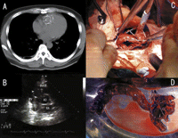
A 38-year-old man was transferred to our hospital with pulmonary embolism. A filter was implanted in his inferior vena cava. Five days later, he had syncope. Computed tomography and transthoracic echocardiography showed the filter in the right ventricle (Fig. 1A–B ). We performed an operation to remove the filter (Fig. 1C–D).

(A) Non-contrast computed tomography showing the filter migrated into the right ventricle (RV). (B) Transthoracic echocardiography demonstrating the filter (arrowhead) in the RV and the right ventricular outflow tract (RVOT). We were not able to perform an endovascular approach to remove filter out of the RV because of a big thrombus (arrow). (C) Intraoperating picture; a thrombus in the RV and the filter that extended into the RVOT. (D) Intraoperating picture; a giant thrombus and the filter removed from the RV and RVOT. This is an extremely rare type of an inferior vena cava filter migration with a giant thrombus, since endovascular approach can be successful in many cases and there is no report of a giant thrombus, such as this case.


