-
PDF
- Split View
-
Views
-
Cite
Cite
Paul E. Achouh, Rachid Zegdi, Arshid Azarine, Jean-Noel Fabiani, Cardiac epicardial tumor with coronary artery compression, European Journal of Cardio-Thoracic Surgery, Volume 35, Issue 2, February 2009, Page 362, https://doi.org/10.1016/j.ejcts.2008.10.044
Close - Share Icon Share
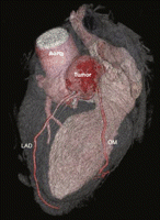
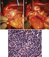
An asymptomatic 58-year-old male was incidentally found to have an epicardial mass on CT scan with compression of left anterior descending coronary artery (Fig. 1 ). Coronary angiogram confirmed compression ( Video 1). Tumor was resected along with left coronary bifurcation (Fig. 2 ). Histopathology showed inflammatory pseudotumor consistent with localized Castleman’s disease. Six-month follow-up CT was normal.

Cardiac CT scan reconstruction showing a round epicardial tumor, 45 mm × 42 mm, located at the level of the proximal left anterior descending artery (LAD) with extrinsic compression of the latter.

(A) Intraoperative view depicting the round epicardial encapsulated tumor, situated just anterior to the left appendage, to the left of the pulmonary artery and at the level of the left coronary artery bifurcation. (B) Intraoperative view showing the tumor dissected out of the cardiac muscle. The proximal LAD was completely inseparable from the tumor. (C) Histopathology view showing a large population of regular plasma cells with hyaline vascular hyperplasia (H&E stain, ×400). OM: obtuse marginal artery; PA: pulmonary artery trunk; Cx: circumflex coronary artery; LA: left appendage; RV: right ventricle.
Appendix A Supplementary data
Supplementary data associated with this article can be found, in the online version, at doi:10.1016/j.ejcts.2008.10.044.


