-
PDF
- Split View
-
Views
-
Cite
Cite
Jinwook Choi, Sungsoo Lee, Jonghwan Moon, Ho Choi, Fish bone induced aortic rupture treated with endovascular stent graft, European Journal of Cardio-Thoracic Surgery, Volume 35, Issue 2, February 2009, Page 360, https://doi.org/10.1016/j.ejcts.2008.10.051
Close - Share Icon Share
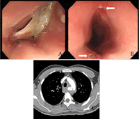
A 31-year-old male presented with fever 3 days after removal of swallowed fish bone with flexible esophagogastroscopy. Multislice CT revealed an aortic rupture (Fig. 1 ). Stent graft was delivered to repair the aortic defect; the esophageal perforation was repaired by early resection and reconstruction with stomach. The patient remains well 4 months after stent graft implantation (Fig. 2 ).

(A and B) Esophagogastroscopy shows the fish bone impacted in the esophagus and esophageal perforation (arrows). (C) Chest CT scan shows contrast leakage from the thoracic aorta.

(A) Intraoperative picture shows the stent graft of aortic rupture (→) and retracted esophagus (↓). (B) Angiography shows the stent graft (32 mm × 10 cm, SEAL Thoracic stent graft, S&G BIOTEC INC.) in the aorta.


