


A 60-year-old gentleman presented with dyspnoea, peripheral oedema and ascites. Radiological imaging demonstrated effusive constrictive pericarditis (Fig. 1 , Video 1). Despite percutaneous drainage of the effusion, cardiac catheterisation confirmed persistence of the constriction with dip-and-plateau ventricular pressure waveform and equalised diastolic pressures. Pericardiectomy, however, resulted in improved symptoms and haemodynamics (Fig. 2 , Video 2).
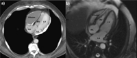
Preoperative computed tomography (a) and dynamic cardiac magnetic resonance images using a steady state free precession sequence (b) demonstrating effusive constrictive pericarditis with a 2 cm anterior pericardial collection compressing the right ventricle and marked calcific thickening of the posterolateral pericardium surrounding the left ventricle. With RA: right atrium, RV: right ventricle, LA: Left atrium; LV: left ventricle.
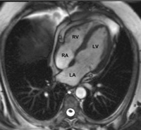
Postoperative dynamic cardiac magnetic resonance images demonstrating reduction of the pericardial collection and improved filling of the right ventricle following pericardiectomy. With RA: right atrium, RV: right ventricle, LA: left atrium, LV: left ventricle.
Appendix A Supplementary data
Supplementary data associated with this article can be found, in the online version, at doi:10.1016/j.ejcts.2008.11.013.
