


A 64-year-old male, whose preoperative 3D-CT revealed arterial bifurcation at the seventh intercostal space (Fig. 1 ), underwent a left lower lobectomy through the same intercostal space for a pulmonary sequestration with a large feeding artery (Fig. 2 ). We found 3D-CT was useful for a safe operation of sequestration with large artery.
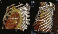
Three-dimensional image obtained from computed tomographic scan revealing a large feeding artery from the descending aorta, as well as a drainage vein to the lower pulmonary vein (A). Bifurcation of the artery from the descending aorta can be seen through the seventh intercostal space (B). Ao: descending artery; FA: feeding artery; ICS: intercostal space.
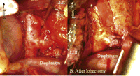
Intraoperative photograph. Large abnormal artery feeding the intralobar sequestration. The anatomical view during surgery was the same as the preoperative 3D-CT imaging. (A) The abnormal artery was clamped with a forceps, then divided and closed with running suture with two pieces of felt (B). S: divided stump of the feeding artery; LUL: left upper lobe; LLL: left lower lobe.
