


A 61-year-old man was presented with a combination of a huge thoracic aortic aneurysm with main pulmonary artery and branch pulmonary arteries dilatation (Fig. 1A and B and Video 1). The patient underwent a cardiac surgery procedure (Fig. 2A and B ). Being very rare, these conditions require special attention and surgical approach.
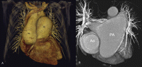
(A) Preoperative 64-slice computed tomography scan demonstrates both ascending aorta (Ao) and pulmonary artery (PA) dilatation. Ascending aorta – 7 × 8 cm in diameter; main pulmonary artery (PA) – 8.1 cm in diameter; right – 3.1 cm; left PA – 6.0 cm; (B) Cross sectional CT image shows both pulmonary artery branches dilatation, with dominant left pulmonary artery ectasia.
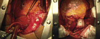
(A) With anulo-aortic ectasia and severe aortic insufficiency, pulmonary artery valve displasia and severe insufficiency, pulmonary artery ectasia, the patient underwent a surgical intervention. Ascending aorta (Ao) and pulmonary artery trunk (PA) were exposed by median sternotomy. (B) Ascending aorta aneurysm and aortic valve were replaced with bioprosthesis and Dacron conduit. Pulmonary artery trunk was open; another bioprosthesis and Dacron tube were used to replace the native valve and pulmonary trunk. Subsequently, the Dacron graft was sheltered by native pulmonary artery wall.
Appendix A Supplementary data
Supplementary data associated with this article can be found, in the online version, at doi:10.1016/j.ejcts.2008.09.045.
