


Abstract
Persistence of the primitive hepatic venous plexus (PPHVP) is an uncommon anomaly that can present associated with complex congenital heart disease and inferior vena cava (IVC) development anomalies and complicate endovascular and surgical procedures. We report a case of PPHVP in a patient with complex congenital heart disease with unexplained persistent arterial desaturation after Kawashima operation and describe magnetic resonance angiography (Angio-MRI) findings leading to the diagnosis. We recommend that IVC-Angio-MRI be performed in preoperative evaluation of patients with complex congenital heart disease and left isomerism when redirection of systemic venous blood to the pulmonary arterial circulation is considered.
1 Introduction
Persistence of the primitive hepatic venous plexus (PPHVP) and portal vein connections is an uncommon anomaly that has been described in patients with interrupted inferior vena cava (IVC) associated with polysplenia syndrome, and also in patients with patent, albeit undeveloped IVC with azygos/hemiazygos continuation to the superior vena cava (SVC) [1–3]. If unrecognized in patients with complex congenital heart disease (CCHD), this anomaly may lead to unexplained profound postoperative hypoxemia due to the diversion of desaturated blood away from the pulmonary arteries with retrograde flow in the azygos vein [1,3]. The angiographic appearance of PPHVP has been previously described [3]. However, the diagnostic findings at magnetic resonance angiography (Angio-MRI) have not been previously reported.
We describe a case of unsuspected PPHVP diagnosed by Angio-MRI in a patient with CCHD presenting with persistent arterial desaturation after the Kawashima operation.
2 Case report
The patient was a 3-year-old girl with a CCHD referred for evaluation with MRI because of development of cyanosis and hypoxemia postsurgery.
The child had been diagnosed with heterotaxy, polysplenia, unbalanced atrioventricular septal defect with hypoplastic left ventricle, bilateral SVCs and interrupted IVC with azygos continuation. She underwent uneventful palliative repair with Norwood intervention and bilateral bidirectional Glenn shunt with azygos incorporation (Kawashima procedure) at 3 weeks and 17 months of age, respectively. In subsequent follow-ups, fatigue, cyanosis and hypoxemia were observed. A cardiac-gated MRI and Angio-MRI confirmed normal-appearing Norwood and bilateral Glenn interventions with patent anastomosis, abdominal situs inversus, interrupted IVC to the right of the aorta with azygous continuation (Fig. 1 ). A striking delay in the delivery of the bolus of contrast material (injected in a peripheral left lower limb vein) to the pulmonary circulation was observed. An early abdominal Angio-MRI venous phase showed a tortuous venous plexus connecting the prerenal IVC to portal venous system and draining to the hepatic veins (Fig. 2 ).
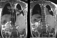
Coronal T2 weighted spin-echo image shows interrupted inferior vena cava with azygos continuation to the right of the abdominal aorta. Note the right-sided gastric camera and spleen.
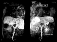
Early phase Angio-MRI with volume rendering 3D reconstruction (VR3D) performed after gadolinium injection in a peripheral left lower limb vein. Contrast material is delivered to the persistent hepatic venous plexus (PHVP) through a spontaneous cavo-portal fistula.
The patient underwent fenestrated Fontan intervention but died during the early postoperative period due to uncontrollable arrhythmia and cardiogenic shock.
3 Discussion
PPHVP has been reported in association with a patent underdeveloped hepatic IVC, CCHD with polysplenia syndrome and azygos/hemiazygos continuation [1–3], and other malformations [1,4,5]. All these cases have been diagnosed by inferior venacavography, with two main patterns [3]. Type 1 consists of a racemose network of middle-sized channels connecting the infrahepatic ICV to the hepatic veins and to the right atrium. Type 2, associated with CCHD with polysplenia syndrome and azygos/hemiazygos continuation of the IVC, consists of a large tortuous venous circuit connecting the infrahepatic IVC and renal vein to the portal system and draining into the hepatic veins through a plexus of intrahepatic venous channels.
In our case, the early venous phase of the abdominal Angio-MRI depicted a tortuous venous plexus arising from the prerenal IVC which connected to the porta vein and drained into the hepatic veins and an interrupted hepatic IVC. The preferential diversion of the venous blood through these channels, may justify the prolonged time in the delivery of the contrast material to the pulmonary circulation observed in our patient, raising suspicion of the presence of a venous anomaly. It has been speculated that the PPHVP is a primary developmental anomaly resulting from the persistence of a primitive hepatic venous network [1,3], supported by its observation in patients with congenital anomalies without previous cavopulmonary surgery [2,3,6]. Most cases of PPHVP were not detected preoperatively because inferior venacavography is not routinely performed before bidirectional cavopulmonary anastomosis. It is important to recognize this venous anomaly for various reasons. Firstly, to avoid the misdiagnosis of IVC stenosis in patients with patent underdeveloped IVC in whom the blood flow is diverted to the PPHVP. Secondly, detection of the PPHVP in patients with left atrial isomerism and CCHD who are being considered for palliative surgery may alter their surgical management. In this setting, coil embolization of the venous fistula before or early after the Kawashima operation, or hepatic vein redirection may be planned. Also PPHVP may complicate or even make unfeasible certain endovascular procedures, such as placement of an umbrella device [4,6].
MRI has an increasing role in the assessment of CCHD, precisely depicting morphological anomalies, and also offering functional information. Angio-MRI provides images of quality comparable to conventional angiography and can demonstrate complex anatomic relationships. In addition, it is non-invasive and avoids ionizing radiation and iodinated contrast-associated toxicity.
PPHVP is an uncommon systemic venous anomaly that can be responsible for persistent arterial desaturation after the Kawashima procedure. Our case highlights the importance of recognizing it before performing a right-to-left shunt surgery. We recommend that IVC-Angio-MRI be performed in routine evaluation of patients with CCHD or left isomerism when surgery to redirect systemic venous blood to the pulmonary arteries is being considered.
