


Several years after an anterior wall infarction, a 62-year-old man experienced a cardiac arrest with successful resuscitation. An internal cardiac defibrillator was implanted. Five years later, echocardiography showed a large thrombus attached to the right ventricle lead (Figs. 1 and 2 ). Thrombus removal, left ventricle remodeling, mitral and tricuspidal annuloplasty were successfully performed.
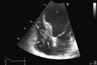
Echocardiography: apical cardiac four-chamber view with the large thrombus (arrows) on the cardiac defibrillator (Prizm VR1850, Guidant Inc.) lead across the tricuspid valve. RA: right atrium; RV: right ventricle: LA: left atrium; LV: left ventricle.
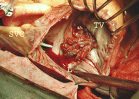
Intraoperative view of the thrombus across the tricuspid valve. Since insertion of the defibrillator, the patient was in sinus rhythm with anticoagulation therapy (fenprocoumon). SVC: superior vena cava; TV: tricuspid valve; L: lead of the cardiac defibrillator.
