


A 44-year-old male with inferior-posterior myocardial ischemia during ECG stress test was referred to 64-slice computed tomography (CT). CT showed a right coronary artery (RCA) fistula draining into the left ventricle (Fig. 1 , Video 1) with dynamic compression of the fistula orifice during systole (Fig. 2 ). CT provides helpful information about fistula location and length before surgery.
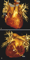
Mega-right coronary artery (RCA) (A) draining into the left ventricle (B) via the posterior myocardium (white arrow) is shown 3D with multislice computed tomography by applying volume-rendering technique (VRT).
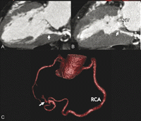
Retrospective ECG-gated image reconstruction by multislice CT showed the fistula open during end-diastole (A) and narrowing of the inner fistula orifice during end-systole (B) due to compression by the myocardium. The inner fistula orifice is located close to the mitral valve (MV) annulus. The RCA had a tortuous ‘8-shaped’ course before its entrance into the left ventricle (white arrow) (C).
Appendix A Supplementary data
Supplementary data associated with this article can be found in the online version at doi:10.1016/j.ejcts.2007.08.027.
