


A 76-year-old female presented with rheumatic mixed mitral valve pathology. A chest X-ray revealed massive cardiomegaly and splaying of the carina (Fig. 1 ). CT scan showed gross isolated left atrial (LA) enlargement (Fig. 2a ), which was plicated from within (Fig. 2b). A mitral valve replacement and LA reduction was performed but she died of an operative complication.
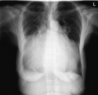
Fig. 1
Chest X-ray shows massive cardiomegaly (cardiothoracic ratio = 1) with splaying of the carina.
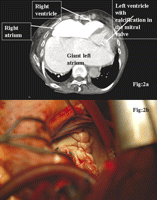
Fig. 2
(a) Contrast-enhanced CT scan shows gross enlargement of the left atrium filling the width of the thorax. (b) Intraoperative picture showing the bulky plication suture line of giant LA appendage from within the left atrium.
European Association for Cardio-Thoracic Surgery
