


A 13-year-old girl presented with cardiogenic shock. Coronary angiography showed an occlusion of the left main coronary trunk (LMT). Enhanced computed tomography revealed a mass plugging in LMT (Fig. 1). The vegetation arising from the aortic wall (Fig. 2) was surgically removed and the aneurismal aortic wall was covered with pericardium patch.
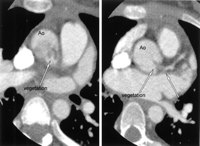
Fig. 1
Enhanced computed tomography revealed a 4×2cm mass in the proximal ascending aorta, plugging in the left coronary main trunk (Ao, ascending aorta; LMT, the left coronary main trunk).
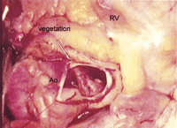
Fig. 2
A friable vegetation (4×2cm) arising from the posterior aneurismal aortic wall was observed (Ao, ascending aorta; RV, right ventricle).
© 2005 Elsevier B.V.
