-
PDF
- Split View
-
Views
-
Cite
Cite
Vassilios P. Argitis, Pierre Schnyder, Ludwig K. von Segesser, Editorial comment: Bauernschmitt et al. “Pitfall in the computed-tomography-diagnosis of postcardiotomy infection: iodine accumulation after irrigation mimicking retrosternal abscess”, European Journal of Cardio-Thoracic Surgery, Volume 27, Issue 4, April 2005, Pages 707–708, https://doi.org/10.1016/j.ejcts.2004.11.029
Close - Share Icon Share
Bauernschmitt and colleagues [1] present an interesting experience, which been underreported until now. As a matter of fact there are numerous reports of mediastinal irrigation with iodine solution for mediastinitis [2–5]. Everybody knows that many contrast media used for angiography or computed tomography are based on water-soluble iodinated solutions.
The authors have to be congratulated for pointing out that post-operative mediastinal irrigation with an iodinated solution must be readily differentiated from an anterior mediastinal infection.
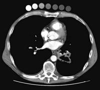
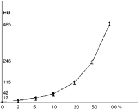
We have looked at this problem in systematic fashion by analyzing CT findings based on various concentrations of iodine solutions, as they are typically used for mediastinal irrigation in post cardiotomy infections, and elsewhere. Solutions of povidone iodine (Betadine®, Mundipharma Medical Company, Basel, Switzerland) with different concentrations 0, 2, 5, 10, 20, 50, and 100% were scanned as shown in Fig. 1, and the correspottnding attenuation values are displayed in Fig. 2. Our investigation clearly shows that povidone iodine at 50% concentration provides a identical contrast attenuation as the contrast enhancement of the aorta.
Post-cardiotomy infection develops in 3% [3] of all patients who had cardiac surgical procedures with extracorporeal circulation and is of major concern to cardiac surgeons. If an aortic graft prosthesis or other implants are present, the problem becomes even more striking due to the difficult management of such complications and their high mortality rates.
A review of the literature reveals that when a radical surgical approach is contraindicated for treatment of mediastinitis, conservative methods can be attempted, with intensive debridement of the infected tissue, local antiseptic irrigation and/or omental transposition [5–7]. Physical examination, clinical and laboratory data as well as CT findings are used to optimize the treatment. The value of CT for the diagnosis of mediastinitis is expressed by a sensitivity of 67% and a specificity of 83% [4].
The use of iodine solutions can improve the validity of CT for the diagnosis of post-cardiotomy infections in terms of sensitivity and specificity. However, CT findings in patients receiving iodine solutions for irrigation are prone to misinterpretation as demonstrated by Bauernschmitt et al. [1]. Hence, in the presence of contrast enhancement at a CT after irrigation, the diagnosis of a water-soluble iodine collection originating from the irrigation has to be ruled out, in order to avoid a false positive diagnosis of an abscess which could lead to an unfounded treatment of a such life-threatening complication.

Seven specimens of Betadine® solutions at concentrations of 100, 50, 20, 10, 5, 2, and 0% (from left to right) have been taped onto the anterior chest wall of a patient prior to an intravenous contrast enhanced helical CT survey of the chest and abdomen for a 5cm excavated carcinoma of the left lower lobe. It appears on this 5mm CT section obtained at 120kV and 120mAs, that Betadine® solution at a concentration of 50% provides an attenuation value of 240 Hounsfield units, which is close to the one in the contrast enhanced lumen of the descending thoracic aorta.

Betadine® concentrations (%) and corresponding attenuation values (HU), as measured on 5mm CT sections obtained at 120kV and 120mAs.


