-
PDF
- Split View
-
Views
-
Cite
Cite
Corinna Ludwig, Ulrike Hoffarth, Jörg Haberstroh, Wolfgang Schuttler, Bernward Passlick, Erich Stoelben, Resistance to pressure of the stump after mechanical stapling or manual suture. An experimental study on sheep main bronchus, European Journal of Cardio-Thoracic Surgery, Volume 27, Issue 4, April 2005, Pages 693–696, https://doi.org/10.1016/j.ejcts.2004.11.030
Close - Share Icon Share
Abstract
Objective: In a previous experimental study on 60 freshly slaughtered pig trachea, a statistically significant better resistance to pressure was found after mechanical stapling compared to hand suture. The objective of this study was to determine the resistance to pressure of a bronchial stump depending upon the closure technique (manual vs. mechanical) used in sheep 14 days after pneumonectomy. Methods: Pneumonectomy was performed on 30 sheep, which were alternatively closed either by a double-layer running suture at 90° to the cartilaginous rings or with an automatic stapling device. Exactly 14 days after pneumonectomy, the animals were sacrificed and the trachea with the bronchial stump was retrieved. Sutures were placed under pressure until air leakage was observed. The air-leakage pressure was recorded digitally. Results: In both groups, there was no evidence of a bronchopleural fistula. As in the previous experimental study, mean values of air-leakage pressure revealed a large standard deviation in both groups (min. 0.16–max. 1.15bar). Unlike the results in the first experiment there was no statistically significant difference between the two groups. Conclusions: After 14 days, when a bronchial stump is considered to be healed, the resistance to pressure of a mechanical suture is equal to that of the manual suture.
1 Introduction
The ideal closure of the bronchial stump after pneumonectomy remains a matter of controversy. Several techniques have been described, as well a variety of suturing materials being proposed to avoid air leakage through the bronchial stump [1–6]. The most serious complication is a bronchopleural fistula with an incidence of 1–10% and a mortality of more than 25% [7–10]. In a large series, Al-Kattan et al. [11] demonstrated that the manual suture of a double layer running suture with polypropylene is efficient. The incidence of bronchopleural fistula was 1.3%. In 1957, the first automatic stapling devices (UKL-60) were used for the ‘en masse’ closure of the pulmonary hilus [12]. Automatic stapling devices have evolved, offering reproducible results and simple rapid handling [13–15]. The development of ‘B’-shaped staples avoids torsion of the staple and permits better neo-vascularisation in the healing bronchial stump. As minimal invasive surgery has become more important in thoracic surgery, the use of automatic stapling devices has increased. Therefore, it is important to know whether one can trust the mechanical suture in the same manner as a manual suture.
In a previous experimental study on freshly slaughtered pig tracheae, we demonstrated that the initial resistance to pressure of the mechanical suture is higher than the manual suture (p=0.011) [16]. The objective of this study was to determine in an animal pneumonectomy model whether after 14 days the resistance to pressure of the mechanical suture remains higher than the manual suture.
2 Materials and methods
2.1 Animal subjects
The study protocol was approved by our Institution's Committee on Investigations involving animal subjects and the Veterinary Department of the local government (G-03/43). All animals were housed and the procedures were performed within the facilities of the University Hospital Freiburg.
2.2 General design
Thirty female sheep with a mean weight of 62.3kg (range; 49–95kg) were randomly divided into two groups. After left-sided pneumonectomy the bronchial stump was closed either with a continuous double-layer running suture or an automatic stapling device. Fourteen days postoperatively, the animals were sacrificed and the main bronchial tree was retrieved. Resistance to pressure was recorded digitally until air leakage occurred and was registered.
2.3 Operative technique
Premedication was performed by the use of 0.05mg/kg atropine (Atropinsulphat Braun®, B. Braun Melsungen, Melsungen, Germany)+0.1mg/kg xylazine (Rompun® 2%, Bayer, Leverkusen, Germany)+20mg/kg ketamine (Ketamin 10%®, Essex, Munich, Germany) intramuscularly. Anaesthesia was introduced through a cephalic vein cannula with 4–6mg/kg propofol (Disoprivan® 1%, AstraZeneca, Wedel, Germany) and after intubation maintained by inhalation of 1.5vol% isofluran (Forene®, Abott, Wiesbaden, Germany) in oxygen/air and additional infusion of 0.1μg/kg/min fentanyl (Fentanyl-Janssen®, Janssen-Cilac, Neuss, Germany). All animals were ventilated with a Servo 900C anaesthesia ventilator (Siemens.Elema, Solna, Sweden). To facilitate pneumonectomy the first suture line of the manual suture for closure of the bronchial stump was performed under apneic conditions, the animals in the manual suture group were preoxygenated (FiO2=1, PaO2≫450mmHg) for at least 6min. Monitoring consisted of online registration of ECG, capnography and blood–gas analysis. Pre-emptive and postoperative analgesia was achieved by administration of 4mg/kg carprofen (Rimadyl®, Pfizer, Karlsruhe, Germany) intravenously and subcutaneously just before the surgical procedure and thereafter every 48h for at least 1 week respectively.
An antimicrobial skin preparation was employed prior to all invasive procedures and was performed in an aseptic surgical manner. Anterolateral thoracotomy on the left side in the fifth intercostal space was followed by pneumonectomy, which was performed in all animals. The bronchial stump was closed with a double layer running suture using Surgilene®—polypropylen 3/0, non-absorbable monofilament, B. Braun-Dexon GmbH, Germany, in apnoea. Care was taken to commence the suture 5–10mm distal to the carina, at the angle between cartilaginous rings and the paries membranaceus on either side with the knot on the outside. The first suture line was a continuous mattress suture placed every 4mm around the cartilaginous rings, knotted at the end. The second suture line was a simple running suture placed every 4mm. Or bronchial closure was achieved with an automatic stapling device using Auto Suture TA 30 Premium® Multifire, Tyco Healthcare GmbH, Germany. Care was taken to place either suture in the ideal position at 90° to the cartilaginous rings. For both methods the time was recorded. The thorax was closed and the animals were extubated.
Fourteen days after pneumonectomy euthanasia was performed with 50mg/kg thiopental i.v. followed by and 2mmol/kg KCL i.v. Through a thoracotomy on the right side the trachea, the right main bronchus and the left bronchial stump was extracted with great care and stored in RPMI 1640 Medium (GIBCO®, Invitrogen Corp., UK) at 4°C until the pressure test could be performed on the same day.
2.4 Material mounting and pressure test
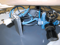
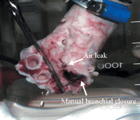
The trachea was affixed to a perforated rubber plug of an appropriate size and then onto the holding device of the testing machine. To avoid the specimen slipping under pressure a wide hose clamp was used. The right main bronchus was closed with a surgical clamp. Air under pressure was slowly applied via a valve through a central perforation of the rubber plug into the lumen of the trachea (Fig. 1). The pressure was slowly increased and recorded digitally until air leakage occurred through the bronchial stump. To facilitate early detection of leakage, the experiment was performed under water (Fig. 2). The maximum pressure at which air leakage occurred was considered as the end-point.
The pressure curve in the bronchial tree was recorded on a monitor (in mbar) using an analogue digital converter to register the data in the computer. Data was documented and processed in Excel (The Microsoft Corp., Redmond, WA, USA). The maximum pressure in the bronchial tree recorded before leakage occurred was determined.
2.5 Statistics
The data was introduced into SPSS (SPSS for Windows, release 11.0, SPSS Inc., Chicago Ill, USA). A descriptive analysis was performed (mean value, standard deviation, minimum and maximum). To test the significance of the difference between the groups, a Mann–Whitney test was performed. The differences were considered significant when the p-value reached ≪0.05.
3 Results
Air leakage through the left bronchial stump was always observed at the bronchial suture. The right bronchial stump was closed with a surgical clamp and showed no air leakage. When air leakage (fall in pressure) was observed the maximum pressure was recorded digitally.
In the manually sutured group, air leakage was mainly observed through the stitches in the paries membranaceus. The air leak in the stapled group occurred at the angle between the cartilaginous rings and the paries membranaceus. In both groups, there was no leakage through the clamped right stump and the suturing material remained intact.
The results for air leakage are documented in Table 1 and Fig. 3. The resistance to pressure of the bronchial stump closed with a stapler was not statistically higher than that of the manual suture (p=0.0663) 14 days after a left sided pneumonectomy.
The time required to close the bronchial stump manually was on average 7.4min (range: 6–10min). The stapled closure took 60s. Considering that in our institution 1min of operating time costs ∼20 Euro and the non-absorbable monofilament ∼3 Euro, the manual suture rounds up to 154 Euro. Comparing this to the stapling device which costs ∼121 Euro, the stapled bronchial closure amounts to a total of 141 Euro.
4 Discussion
To avoid a bronchopleural fistula, several closure techniques have been proposed and discussed. The quality of the double layer running suture was proven by Al-Kattan et al. [11], with a very low bronchial stump insufficiency rate in a large series. Ever since the introduction of automatic stapling devices, they have been compared with manual suturing [9,13,14,17,18].
Even a cough can produce a considerable rise in pressure, up to 200mmHg in a young man [19], leading to air leakage if the bronchial stump is insufficient. The objective of this study was to focus on one single factor in a previously established model [16], under ideal circumstances, once the bronchial stump is considered to be stable (healed): the resistance to pressure. A device was developed to increase pressure within the bronchial stump until air leakage occurred, which was registered digitally. Other factors influencing the stability of bronchial stump such as, age, vascular status of the patient, extent of lymphadenectomy, pre- and post-treatment with radiation and chemotherapy, bacterial infection, and length of the bronchial stump, were not considered.
In contrast, to the results obtained in our previous study on freshly slaughtered pig tracheae where the mechanical suture was more resistant to pressure [16], 14 days after pneumonectomy, under ideal conditions, there is no longer any difference in favour of the automatic stapling device. The higher stability and therefore the increased resistance to pressure of the mechanical suture disappears once the bronchial stump has healed. Similarly to the previous results, a high variability within both groups was found (Table 1). This can be attributed to the individual properties of the bronchial wall.
If the stability of the bronchial closure is equivalent after 14 days then the cost of the material may be important for the decision to close the bronchial stump either by hand suture or a mechanical stapling device. Since, the closure of the bronchus by hand suture is much more time consuming as compared to the mechanical stapling of the bronchus (1 vs. 7.4min), stapling seems to be more cost effective. Furthermore, application of the mechanical stapling device avoids contamination of the thoracic cavity by tracheal and bronchial secretions.
In conclusion, in an in vivo model of pneumonectomy there is no difference with respect to resistance to pressure of the bronchial stump when comparing mechanical stapling with manual suture under ideal circumstances. However, this might not be true for ‘critical circumstances’ such as poor blood supply, infection or after radio-chemotherapy. This has to be evaluated in additional studies.

A mechanical device with a perforated rubber plug onto which the trachea is mounted and through which the pressure is applied onto the bronchial suture.

Air leak through the paries membranaceus of the bronchial stump with manual suture.

Box plot of the mean values and the standard deviation for the maximum pressure in the two groups.


