-
PDF
- Split View
-
Views
-
Cite
Cite
Witold Rzyman, Jan Skokowski, Radosław Wilimski, Krzysztof Kurowski, Mirosław Stempniewicz, One step removal of dumb-bell tumors by postero-lateral thoracotomy and extended foraminectomy, European Journal of Cardio-Thoracic Surgery, Volume 25, Issue 4, April 2004, Pages 509–514, https://doi.org/10.1016/j.ejcts.2003.12.022
Close - Share Icon Share
Abstract
Objective: Thoracic dumb-bell tumors are rare, usually benign tumors in the posterior mediastinum, consisting of intrathoracic and intraspinal parts. Surgical removal is the treatment of choice, performed by two teams – neurosurgeons and thoracic surgeons operating in a prescribed order. Methods: Between 1994 and 1997 five patients had dumb-bell tumors removed in a one-step operation involving postero-lateral thoracotomy and extended foraminectomy. This operating method, rarely described in the medical literature, consists of intrathoracic and intraspinal parts being performed by a thoracic team independently or with the assistance of a neurosurgeon. Initially the intrathoracic part is resected, followed by an extensive widening of the intervertebral foramen to an appropriate extension and the removal of the remaining intraspinal part of the tumor. Results: Four postero-lateral thoracotomies and one incision over a huge tumor in the thoraco-lumbal region, without entering the pleural cavity, were performed. In one patient postoperative, transient leakage of the cerebral fluid was observed. No form of late complications or neurologic sequelae have been reported within a 5-year follow-up. Conclusions: One-step removal of a dumb-bell tumor by postero-lateral thoracotomy and extended foraminectomy is a safe surgical procedure that can be performed by the thoracic team alone. Early and late surgical results confirm the appropriateness and usefulness of the method.
1 Introduction
Tumors of the posterior mediastinum arising from the nervous system and penetrating through the intervertebral foramen into the spinal canal are called dumb-bell or sandglass tumors [1]. In the majority of cases they are neurogenic benign tumors which have their origin in the neuron sheath (68%), parasympathic ganglion (30%) or paraganglionic cells (2%) [2]. Only 3–19% of such tumors, usually in children, are malignant. Akawari [1] reported that 10% of all the mediastinal tumors of neurogenic origin extended from the thorax through the spinal foramen into the neural canal. The intraforaminal growth may cause destruction of the vertebral body, while its intraspinal component may cause spinal cord damage by compression or when malignant infiltration with neurological sequelae [3]. Weber in 1856 was the first scientist to describe such tumors but, in 1952, Love and Dodge [4] first introduced the name ‘dumb-bell’ tumors reporting the ingrowths of a neurogenic tumor into the spinal canal. Patients usually present neurological symptoms but some asymptomatic dumb-bell tumors have appeared during a routine chest X-ray [3].
We present a method of removing thoracic dumb-bell tumors in a one-step operation through postero-lateral thoracotomy and extensive widening of the intravertebral foramen—foraminectomy. Our experience regarding the removal of dumb-bell tumors presented in this report is based on five patients operated during the period from 1994 to 1997. In all of these cases we applied a one-stage removal by postero-lateral thoracotomy and extended foraminectomy. A team of thoracic surgeons operated on all the patients assisted by a neurosurgeon both at the beginning and whenever the extension of the intraspinal part was expected to be widespread.
2 Materials and methods
Between 1994 and 1997 five patients were admitted for a thoracic dumb-bell tumor removal at the Department of Thoracic Surgery in the Medical University of Gdańsk. Three men and two women, between 35 and 70 years old (mean age of 51) were operated on by applying foraminectomy to remove the intraspinal parts of the tumors. In four of these cases postero-lateral thoracotomy was performed and in the remaining one patient, an incision over the tumor in thoraco-lumbal region. Four patients had symptoms of the spinal compression, while one was asymptomatic and the tumor mass was diagnosed coincidentally. During a preoperative assessment only patient no. 3 had a fine needle biopsy with the diagnosis of a benign neural tumor. All of these patients before being admitted into the Department of Thoracic Surgery had undergone computed tomography (CT) scans and two of them, in addition, magnetic resonance imaging (MRI) scans of the thorax (Figs. 1 and 2) . The diagnostic protocol for the patient with dumb-bell tumor in our institution includes chest radiograph, CT thorax and neurological examination. If intraspinal tumor extension is significant and/or infiltration of the surrounding structures including spinal cord is suspected, MRI and fine needle biopsy is performed.
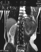
Magnetic resonance imaging of the thoracic dumb-bell tumor—axial reconstruction (patient no. 3).
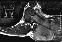
Magnetic resonance imaging of the thoracic dumb-bell tumor—normal scan (patient no. 3).
Patients were followed up in our outpatient clinic every 3 months in the first year and then once a year after the operation. CT scans were performed in two of them postoperatively.
2.1 Description of the operation
The patient is placed on the side in the lateral decubitus position. Postero-lateral thoracotomy is performed through the intercostal space corresponding with the level of tumor penetration to the spinal canal.
After the opening of the pleural cavity, the lung is moved forward to reveal the intrathoracic neurogenic mass. The parietal pleura over the tumor is incised and detached. The dissection of the tumor extends into the intervertebral foramen. At this stage, the decision is made whether the intrathoracic part of the tumor should be removed or left until the resection of the tumor's intraspinal part is complete. If the manipulation of the tumor is difficult and the view into the intravertebral foramen insufficient we either cut the intrathoracic part off completely or we leave only a small portion of it to be held with the artery forceps. Now, blunt and sharp dissections in the proximity of the foramen permit the lifting of the tumor, exposing the narrowed part placed in the foramen. The tumor is gradually dissected off its surroundings, while being lifted up by hand or the artery forceps. This maneuver affords a good view into the foramen. When the tumor has a wide penetration to the neural canal, one can begin to widen the foramen in a chosen direction: upwards, downwards or both. If necessary, the processus tranversus is removed allowing a better view into the structures of the spinal canal (Fig. 3) . After the opening of the vertebral canal, one proceeds with a very delicate dissection, with either the scissors or coagulation, of the remaining tumor tissue from the dura mater. In case of a hemorrhage, coagulation, hydroxide peroxide or a sponge were used. No sealing material was applied to fill the opening after foraminectomy.
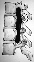
Extended foraminectomy. The opening connecting three foramina. Dashed line around the processus transversus illustrates possibility of its removal.
3 Results
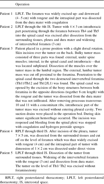
The details concerning the patients' symptoms, preoperative radiological and postoperative pathological findings are listed in Table 1 , while a description of each patients' operation has been included in Table 2 . Four of the five tumors gave symptoms usually associated with pressure on the surrounding structures. Three patients had no findings in a neurological examination. The CT was done in all patients and demonstrated intraforaminal growth of the tumor and its intraspinal extension. When infiltration of the surrounding structures including spinal cord was considered after CT, MRI of suspected region was employed. In four patients postero-lateral thoracotomy and in one (patient no. 3) an incision over the tumor at the back, without entering the pleura itself, was performed. Three of the tumors had a wide intraspinal part, it was smaller in the case of the remaining two, requiring only a 3-cm widening of the foramen. In two patients an opening of 4–5 cm was enough, but in patient no. 3 a length of 8 cm along the lateral part of the vertebral body between two foramina (Th11/12 and Th12/L1)—that extended additionally in the opposite directions—had to be excised (Fig. 3).
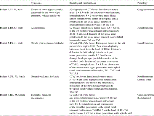
Symptoms, radiological findings and histopathological diagnosis in all patients
In one patient with a huge tumor in the lowest part of the thoracic cavity, intraoperative bleeding from the venous plexus of the spinal canal followed by a leakage of the cerebral fluid, occurred postoperatively. He had neurological sequelae in the form of transient meningitis-like symptoms (4 days) with a minimal Babinski symptom on the left side, a bilateral Kernig symptom and neck stiffness. All were successfully treated with bed rest and fluid balance in 7 days. All of the patients are monitored once a year in an outpatient clinic, and no late neurological sequelae have been noted.
4 Discussion
A surgical excision of the dumb-bell tumor, which is the treatment of choice, is a challenge to the thoracic surgeon mainly because it covers two surgical fields: thoracic and neurosurgical. Therefore surgical techniques and approaches vary widely. Some surgeons, depending on the institution, prefer a two-stage operation, where the removal of the intrathoracic and intraspinal tumor mass is done separately and in different sequences. In the late 1970s, however, some surgeons introduced a one-stage removal of the dumb-bell tumor. Akawari, for example, combined two approaches in a single session. The first step was posterior laminectomy, done by neurosurgeons, and the second step was postero-lateral thoracotomy, performed by a thoracic team. This method, in the opinion of the authors, avoids the risk of bleeding from remnant tumor tissue, compression of the spinal cord, leakage of the cerebral fluid or damage to the spinal cord encountered in a two-stage procedure [1,3]. Grillo, in 1983, demonstrated a one-approach technique, i.e. through a band skin incision at the back. The first step consisted of removal by a neurosurgeon of the intraspinal part by laminectomy, with the rest of the tumor being removed by a thoracic surgeon through thoracotomy [3,8,9]. The only difference between these two methods is the skin incision. Ricci et al. [10] concluded that Grillos method only allows the removal of dumb-bell tumors whose intraspinal part does not exceed 3 cm. Lately, a combined approach involving laminectomy by a neurosurgeon followed by a videothoracoscopic removal of the intrathoracic part has been the focus of interest [11–14].
In a preoperative diagnosis, a fine needle biopsy is rarely successful due to the consistency (hard and solid) and location (aorta, vertebral structures) of neurogenic tumors. Since almost all such tumors are benign in adults [1,5] and they need to be removed anyway, it is performed on very rare occasions. Nor is the use of MRI obligatory in the diagnosis, in our opinion. Its advantage over a CT scan lies mostly in the accuracy of describing the longitudinal extension of the intraspinal component of the tumor [10]. Either method is capable of visualizing the morphologic features of the neural mass, but infiltration of soft tissues including neural structures is better seen on MRI images. Although Shadmehr et al. [9] reports that CT missed to show intraforaminal growth in three of 16 reported cases in their series we have never been surprised by such accidental findings while operating neurogenic tumors at our institution. Myelography is only of historical significance [15]. Arteriography is rarely, if ever necessary, and maybe helpful when considering proximity of Adamkiewicz artery in the lower portion of the posterior mediastinum, infiltration of aorta or major aortic branches [9]. Although the majority of dumb-bell tumors are of benign origin, careful diagnosis with CT, MRI, or biopsy is mandatory in the rare cases of their being malignant.
Dumb-bell tumors of the thoracic region comprise 10% of all the neurogenic mass in the posterior mediastinum [1,5]. Most of the neurinomas are easy to remove by subtle dissection in the proximity of the foramen followed by slow and deliberate extraction of the tumor from the spinal canal. In the majority of ‘true’ dumb-bell tumors, however, a good view over the medulla is necessary. In such cases, postero-lateral thoracotomy with widening of the intervertebral foramen by the resection of its bony structure is preferable [6]. If necessary, the processus transversus can be excised, affording an excellent overview of the medulla. Five patients have been operated on in this manner and subjected to long-term follow up. We have been able to find in the literature only one case report concerning the removal of dumb-bell tumor in the cervical region by foraminectomy [7].
Our technique of one stage dumb-bell tumor resection through postero-lateral thoracotomy and extended foraminectomy can be regarded as a safe procedure leading to satisfying early and long-term results without neurological sequelae. The intervertebral foramen could be excised very extensively. In patient no. 3 the extensions of the opening exceed the distance between two foramina. We find the operative technique easy in the hands of an experienced thoracic surgeon who has good control over the tumor and the surrounded structures at all stages of the operation. Earlier on, we used to ask neurosurgeons for assistance during the intraspinal part of the operation, but in the last three patients the thoracic team alone performed the operation. When the tumor is malignant, naturally, it is preferable to have a neurosurgeon in the operation room while operating dumb-bell tumor with this technique. Absence of instability of the spine or neurologic symptoms of neural compression or destruction confirms, however, the usefulness and effectiveness of the applied operative method. In two operated patients we have used gelatin sponges (Surgicel and Spongostan) to stop bleeding from the resected foramen. Due to recent case reports of paraplegia caused by swelled sponges in the postoperative period we advise to avoid application of similar materials or to use them with appropriate caution [16,17]. An interesting alternative to our technique is a dorsal approach, proposed by Osada et al. [2], involving laminectomy and resection of a small portion of the adjacent rib without opening the parietal pleura. The avoidance of thoracotomy helps in postoperative pain control.
Due to limited series of dumb-bell tumors with wide extension in the spinal canal no institution sees a lot of such neoplasms. Therefore, the appropriate method should be applied concerning the variety of anatomic situation and experience of the operating team. In our opinion extended foraminectomy has its place in surgical treatment of thoracic dumb-bell tumors but it should be performed by highly experienced thoracic surgeons.


